The content on this page is intended to healthcare professionals and equivalents.
Today, diseases derived from lifestyles are increasing worldwide.
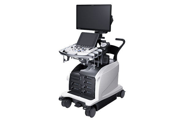
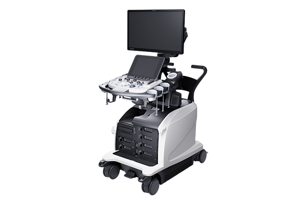
ARIETTA 750SE provides advanced diagnostic information quickly with various functions and advanced applications that support a smooth workflow.
Prior fixed examination protocols and imaging conditions can be registered. Button operations can be reduced significantly to support efficient examinations. Additionally, a reference image can be displayed via the "Guide View" function.


HI Strain is an algorithm used to display an Elastography image more consistently than before. It is possible to display Elastography images with high continuity while maintaining temporal resolution and spatial resolution.
A series of operations from frame selection to the boot of FLR measurements are automated. Elastography evaluation with high reproducibility is possible via automation.

Automation of Measurement
RTE assesses tissue strain in real time and displays the tissue stiffness as a color map. Its application has been validated in a wide variety of clinical fields, and it is possible to calculate an estimate value of liver fibrosis staging.
It is possible to evaluate tissue stiffness by generating shear waves and measuring Vs, its propagation velocity in the tissue. Combi-Elasto, which integrated RTE and SWM, is expected to be used for cases which are difficult to diagnose through only using SWM.
A function to measure the attenuation coefficient generated in the process of ultrasound propagation of tissue. The degree of steatosis can be estimated from the size of the coefficient. Its measurement is conducted simultaneously with SWM, and it can be conducted as an extension of B mode examination.

Contrast enhanced ultrasound is used widely for clinical practices such as tumor detection, differential diagnosis, and treatment support. High definition and high sensitivity contrast imaging is realized by Variable Beamformer and high sensitivity transducers.
Imaging technology for visualization of low velocity blood flow below the previous detection threshold. A unique algorithm displays fine blood flow with greater resolution and sensitivity.

MPR images constructed from CT/MRI/PET-CT volume data can be synchronized to real-time ultrasound imaging. It is applied in a wide variety of clinical fields: such as for Abdomen, Breast, navigation in prostate puncture, and so on.