


The content on this page is intended to healthcare professionals and equivalents.
The advanced imaging technolgy for highly dynamic visualisation of low velocity blood flow below the previous detection threshold with high frame rate. The algorithm displays a clear and accurate information on blood perfusion with greater resolution and sensitivity.
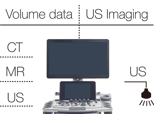
MPR images constructed from CT/MR/US volume data can be synchronized to real-time ultrasound imaging. It allows a direct comparison of the lesion depicted by each imaging modality, taking advantage of the different imaging methods.
RTE assesses tissue strain in real time and displays the measured differences in tissue stiffness as a color map. Its application has been validated in a wide variety of clinical fields: for the breast, thyroid gland and urinary structures.
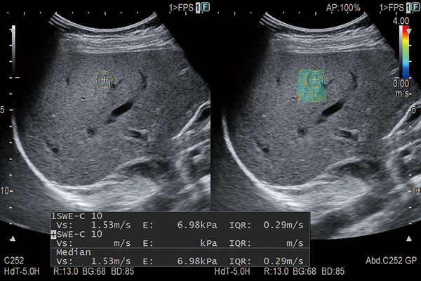
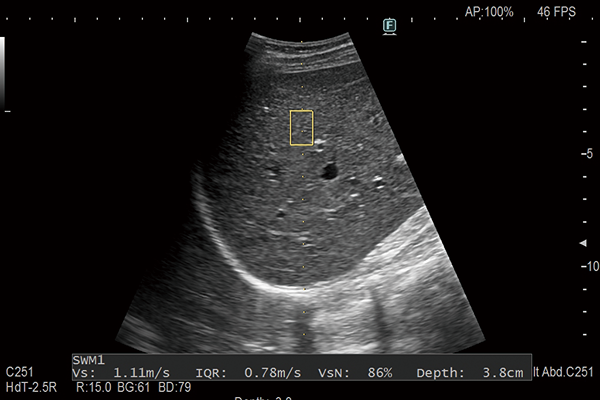
Shear waves are generated using a ‘push pulse’ to excite the tissues. SWM provides an assessment of tissue stiffness by calculating Vs, the propagation velocity of the shear waves. SWM provides an additional reliability indicator, VsN, as an objective evaluation of the Vs measurement. SWE color-codes tissue sttifness based on the propagation velocity of shear waves. SWE can be used to evaluate liver visually and non-invasively.
iATT non-invasively measures the attenuation of ultrasound to provide information for evaluating the amount of fat in the liver. Easy ROI setting achieves to reduce measurement influence from multiple reflections and structures such as blood vessels by narrowing the analysis area and guide display.
Three- and four-dimensional imaging can play a role as a prenatal communication tool connecting parents with their fetus. Auto Clipper automatically defines the optimal cut plane removing placental or other unwanted tissue signals in front of the fetus, offering a clear surface-rendered fetal image.
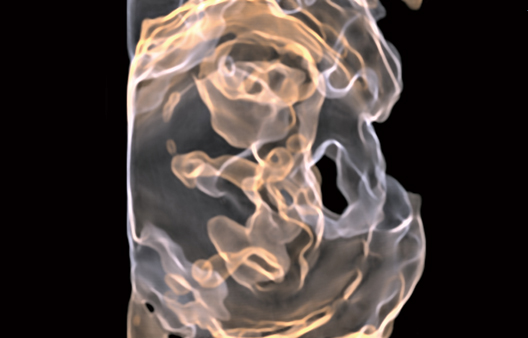
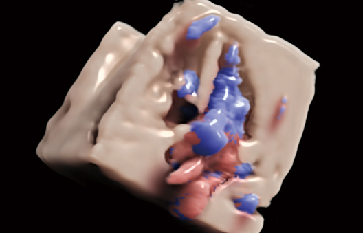
The 4Dshading technology gives a more realistic appearance to the rendered surface of the fetus in the 3D display. 4Dshading Flow is its Doppler blood flow mode tuned to offer a better understanding of complex vascular flows. 4Dtranslucence enables evaluation of fetal structures providing a display of the fetal body surface and internal organ boundaries with a translucency.
Automatic tracking of fetal heart movement from the B mode image follows the displacement of the heart wall in the apical direction for measurement of %Fractional Shortening (%FS). Measurement is unaffected by a change in the fetal position or by the mother’s breathing.
Enables observation of Doppler waveforms from two different locations during the same heart cycle. Simple measurements from two different waveforms can also be useful in the diagnosis.
- *1 Option