


The content on this page is intended to healthcare professionals and equivalents.
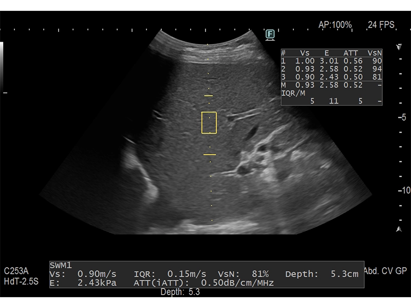
SWM assesses tissue stiffness non-invasively by calculating Vs, the shear wave propagation velocity. iATT measures ATT (Attenuation) index to estimate the degree of hepatic steatosis. SWM and iATT can be measured simultaneously, allowing for simple and convenient operation.
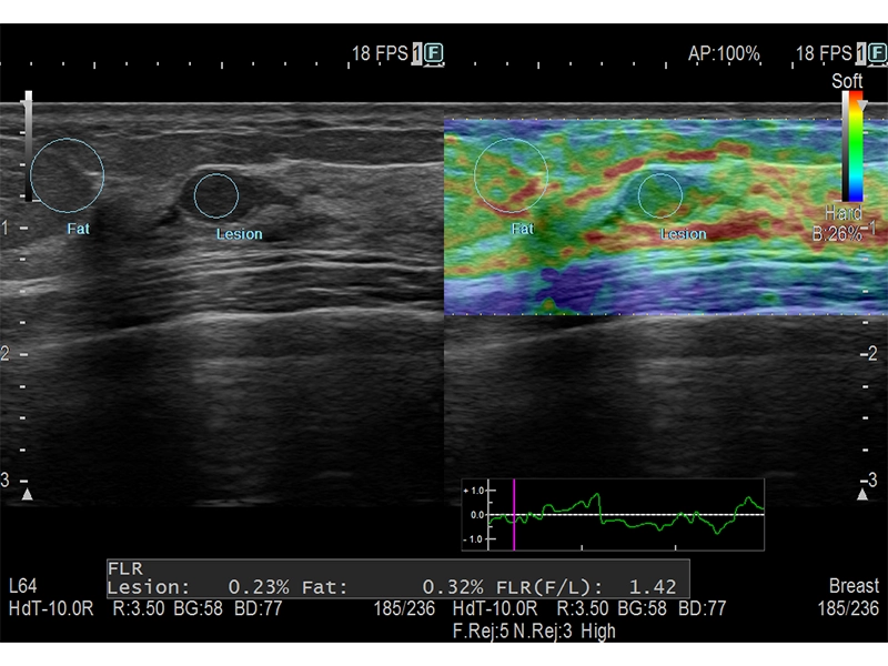
Assesses tissue strain in real time and displays the measured differences in tissue stiffness as a color map. In breast imaging, a series of operations—from selecting the appropriate measurement frame to initiating FLR (Fat Legion Ratio) analysis—can be performed automatically, enabling easy and reproducible evaluations.
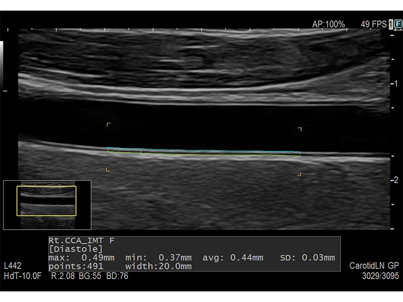
By simply setting an ROI (Region of Interest) on the longitudinal B-mode image of the carotid artery, max IMT and mean IMT are automatically measured. Because maximum, minimum, mean, and standard deviation values are calculated from all points within the specified range, this method is expected to offer a more objective assessment compared to manual measurement.
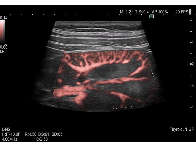
DFI visualizes fine blood flows with high resolution and sensitivity. Additionally, it features effective noise reduction capabilities caused by transducer scanning or patient movement, enabling stable observation of blood flows.
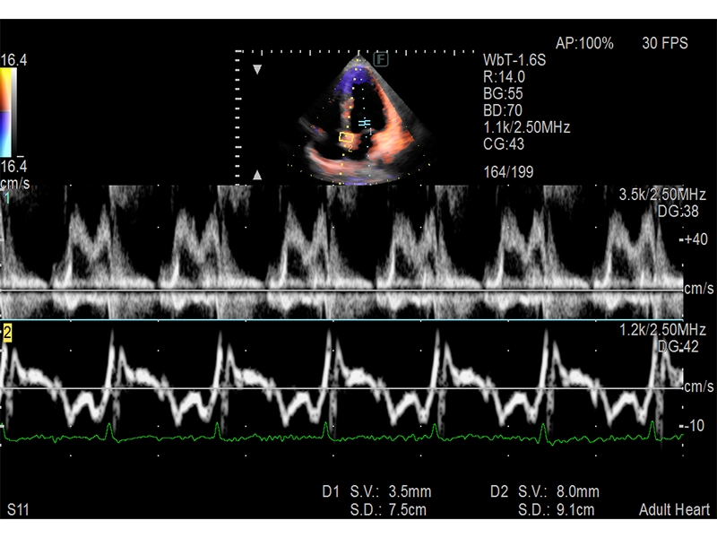
It can observe Doppler waveforms at two different sites simultaneously within the same heartbeat and phase. Combinations of PW/PW, TDI/TDI, and PW/TDI* are possible. Measurements such as E/e’, an indicator of left ventricular diastolic function, can be performed at the same phase even in cases with arrhythmia.
- * Depends on the transducer used
- *1 Option