France

Women's Health

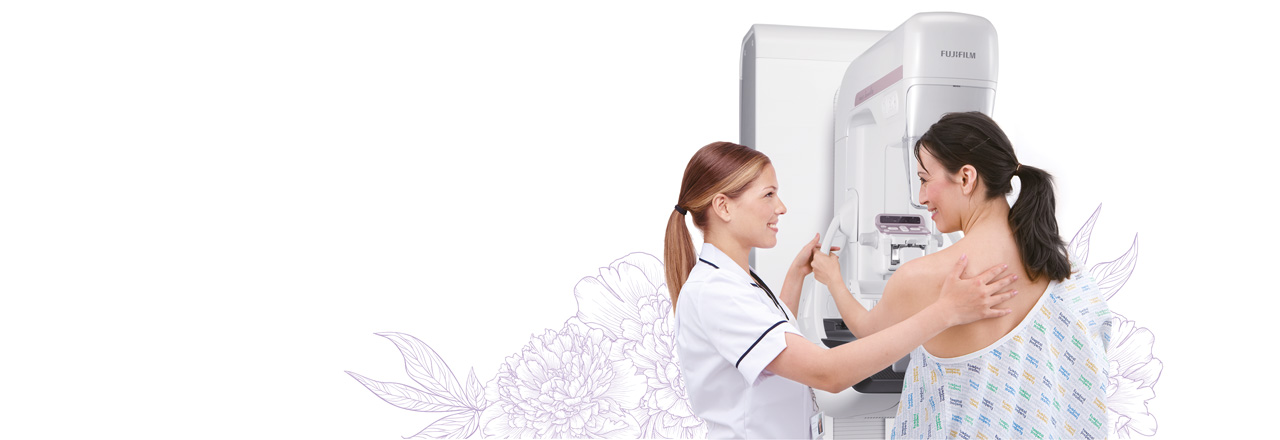
With Amulet Innovality, we are committed to clinical excellence in acquisition and diagnostic reading, with the woman at the center.

The content on this page is intended to healthcare professionals and equivalents.

The Innovality mammography system is the expression of technology evolution in breast imaging made possible by Fujifilm’s commitment in continuous research and development.

“The “Harmony” version of the AMULET Innovality mammography system improves the instruments that allow you to work on precision images combining Fujifilm’s advanced technologies, Dynamic Visualization II and Iterative Super Reconstruction, improving reading comfort and speed due to balanced image processing and display of all relevant details in one image[1] said Eiji Ogawa, VP Modality Solutions, QA and RA FUJIFILM Europe GmbH “In addition, improved S-View offers a new synthetic 2D image that combines 3D power and 2D reading speed. These innovative features help the doctor’s diagnosis, aiming to make it more accurate and precise.”

Technical Specifications

Main Unit

Main unit

The Innovality mammography systems represents Fujifilm’s commitment in radiology equipment development and manufacturing: its design embedded with proprietary technologies and components, makes it the proud flagship of our digital mammography portfolio.
Tube and generator

The  Innovality mammography system features a tungsten anode tube, alternatively coupled with three different filters:

Rhodium filter for an optimized beam for 2D exposures;
Aluminium filter to be used in tomosynthesis exposures;
Copper filter to be used for energy subtraction exposures (CEDM)
Detector

The  Innovality mammography system employs a direct-conversion flat panel detector made of Amorphous Selenium (a-Se) which exhibits conversion efficiency in the mammographic X-ray spectrum.

The new HCP (Hexagonal Close Pattern) detector efficiently collects electrical signals converted from X-rays to realize both high resolution and low noise. This design makes it possible to realize a higher DQE (Detective Quantum Efficiency) than with the square pixel array of conventional TFT panels. With the information collected by the HCP detector, the AMULET Innovality mammography system creates high definition images with a pixel size of 50 μm.

Intelligent AEC optimizes the X-ray dose for each breast type

Intelligent AEC has advantages in defining the adequate dose for an examination compared to conventional AEC systems where the sensor position is fixed. Through the analysis of information obtained from low- dose preshot images, Intelligent AEC makes it possible to consider the mammary gland density (breast type) when defining the x-ray energy and level of dose required. Able to be used even in the presence of implants; intelligent AEC enables more accurate calculation of exposure parameters than is possible with conventional AEC systems. By allowing the use of automatic exposure for the implanted breast, Intelligent AEC can further enhance examination workflow.

Patient information display

The information shown on the display at the base of the exposure unit can be switched between patient information (ID, name, date of birth, etc.) and positioning information (angle of swivel arm, compression force and breast thickness). Positioning information can also be confirmed on the display on the compression arm.

Patient comfort

The AMULET Innovality mammography system has been designed with patient comfort in mind, and can reduce the discomfort which is typical of mammography exams.
The gantry design embeds two wide and easily accessible armrests and offers the possibility to install an additional lower one (to be positioned under the detector).

Acquisition Workstation

Completely integrated with the main unit, the AMULET Innovality acquisition workstation (AWS) is the “Central Hub” of operation: it is a multi-purpose tool, that allows efficient and seamless planning and verification of each step in digital mammography workflow:

Integrated X-ray controller allows setting and confirmation of exposure conditions on a single screen.
Examination screen can be split and switched between 1, 2, or 4 image display.
Individual images can be immediately output to a PACS, viewer or printer during an examination.
Density and contrast can be easily adjusted while viewing images.
Alignment of left and right images can be adjusted both automatically and manually.

Dual display configuration

A second, high resolution monitor can be added to the AWS making it possible to display previous images recalled from a PACS to ensure the operator has access to previous images at all times.
For Tomosynthesis, reconstructed images can be displayed.

Variable height desk

A new desk has been designed to improve operators comfort and system ergonomics, which is featured with an automated adjustable height mechanism: each user is able to operate in the best conditions and safety.
The desk is not available outside of Europe.

Tomosynthesis

In breast tomosynthesis, the X-ray tube moves through an arc while acquiring a series of low-dose X-ray images. The images taken from different angles are reconstructed into a range of Tomosynthesis slices where the structure of interest is always in focus.


The reconstructed tomographic images make it easier to identify lesions which might be difficult to visualize in routine mammography because of the presence of overlapping breast structures.
The Tomosynthesis function on AMULET Innovality is suitable for a wide range of uses, offering two modes to cater for various clinical scenarios. Standard (ST) mode combines rapid exposure timing and efficient workflow with a low X-ray dose while High Resolution (HR) mode makes it possible to produce images with a high level of detail, allowing the region of interest to be brought into clearer focus.

Routine mammography and breast screening: ST MODE

Acquisition angle: ±7.5°
Pixel size: 100/150 μm
The smaller angular range and fast image acquisition allow Tomosynthesis scans to be quickly performed with a relatively low x-ray dose.

The high acquisition efficiency provided by ST mode has been designed to allow routine Tomosynthesis application, supporting diagnostic mammography on asymptomatic patients and its implementation in breast screening.

Diagnostic mammography: HR MODE

  • Acquisition angle: ±20°
  • Pixel size: 100/50 μm

With a larger acquisition angle the depth resolution is improved. This allows the region of interest to be defined more clearly and brought into clearer focus.

HR mode allows tomosynthesis acquisition to reach higher detection accuracy: details depiction and structure conspicuity are supported by a higher spatial resolution and a more efficient overlaps removal. The ideal tool to investigate suspicious findings or to evaluate symptomatic women.

Image processing

ISC Image-based Spectrum Conversion

Biopsy

The AMULET Innovality mammography system features advanced tools to support biopsy procedures.

Seamless and integrated workflow, automatic and mechanical needle positioner, flexible configuration and setup: a tool to bring interventional procedures to a higher efficiency and accuracy level.


Tomoguided targeting (tomobiopsy)

“The “Harmony” version of the AMULET Innovality mammography system improves the instruments that allow you to work on precision images combining Fujifilm’s advanced technologies, Dynamic Visualization II and Iterative Super Reconstruction, improving reading comfort and speed due to balanced image processing and display of all relevant details in one image[1] said Eiji Ogawa, VP Modality Solutions, QA and RA FUJIFILM Europe GmbH “In addition, improved S-View offers a new synthetic 2D image that combines 3D power and 2D reading speed. These innovative features help the doctor’s diagnosis, aiming to make it more accurate and precise.”

Stereotactic targeting

The stereotactic biopsy unit allows accurate and reliable biopsy procedures to be performed using 50μm pixel images: not even a subtle detail is missed for extremely accurate targeting.

Operating display

Together with the biopsy positioner, an additional operating display is being attached to the gantry: automatic operation can be easily started using this interface, as well as any manual adjustment or override.

Lateral Needle Holder

Fujifilm’s biopsy positioner allows multiple combination between gantry orientation and needle approach: thanks to a specific adapter the lesion can be reached either following a vertical and conventional orientation (perpendicular to detector) or with a lateral approach (parallel to detector).

Compression Paddles

CONTINUOUS INNOVATION IN DIGITAL MAMMOGRAPHY

Commitment to Women’s health in everything we do, from providing educational programs to ensuring a comfortable and safe mammography procedure.  

EXPERIENCING FUJIFILM DIGITAL MAMMOGRAPHY

Learn the most recent experiences from Fujifilm Key Opinion Leaders.

Download case studies and technology reviews from the "Take Away" series.