


The content on this page is intended to healthcare professionals and equivalents.
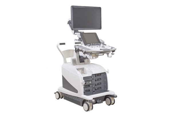
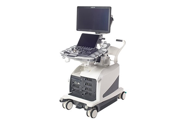
The ARIETTA 850 DeepInsight has advanced applications that quickly provides diagnostic information keeping it one step ahead of the market.
RTE assesses tissue strain in real time and displays the measured differences in tissue stiffness as a color map. Its application has been validated in a wide variety of clinical fields: for the breast, thyroid gland, and urinary structures.
It is possible to evaluate tissue stiffness by generating shear waves and measuring Vs, its propagation velocity in the tissue.
An index to estimate the degree of hepatic adipogenesis (ATT) can also be measured at the same time.
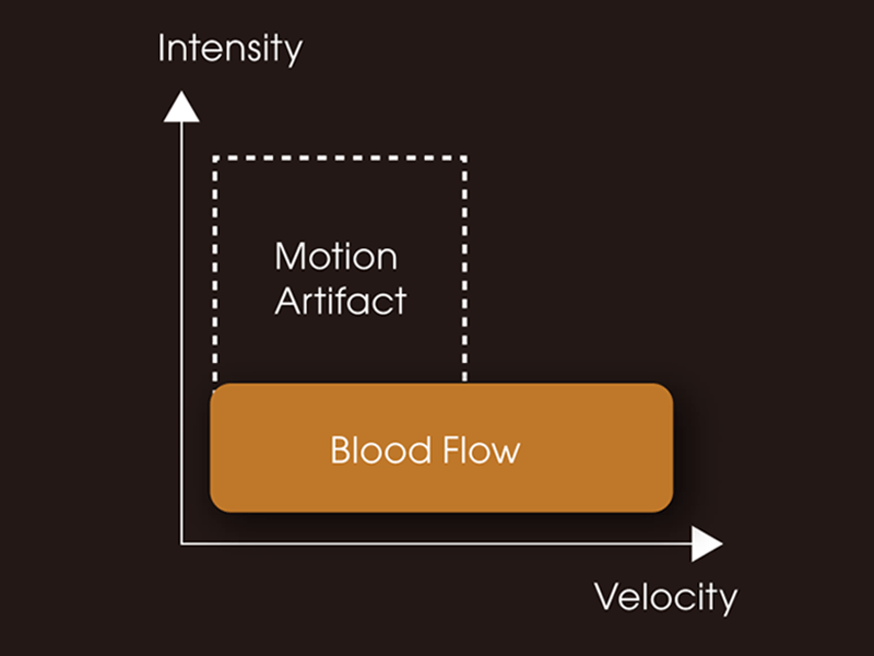
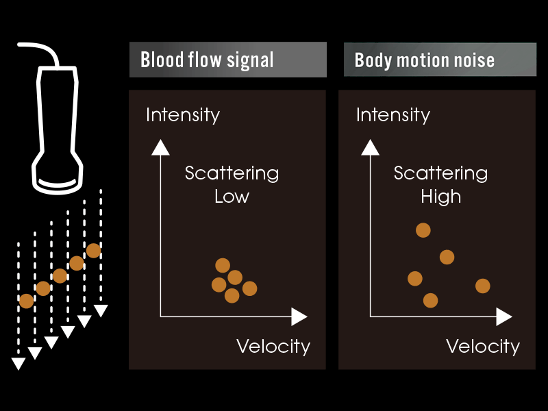
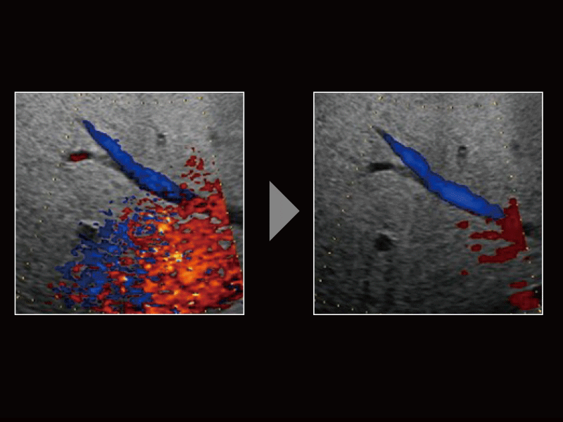
Detective Flow Imaging (DFI), which is a micro vascular flow imaging technology, has supported noise processing technology “Wall Motion Reduction PLUS” so that body motion noises are suppressed selectively, and images of high sensitivity and visibility can be realized. “Wall Motion Reduction PLUS” judges the scattering of detected signal intensity to perform noise reduction according to the body motion amount of each region.
DFI
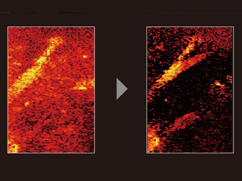
Judge the scattering of signal intensity to reduce noise
A wide variety of transducers and advanced functions support a treatment.
-Realize higher sensitivity by filter processing improvement and signal velocity scattering judgement-
Flow sensitivity is improved by shifting the wall filter to the low velocity side. With the filter processing changed, body motion noises are reduced effectively by detecting the signal velocity scattering in multiple-times receiving and judging them.
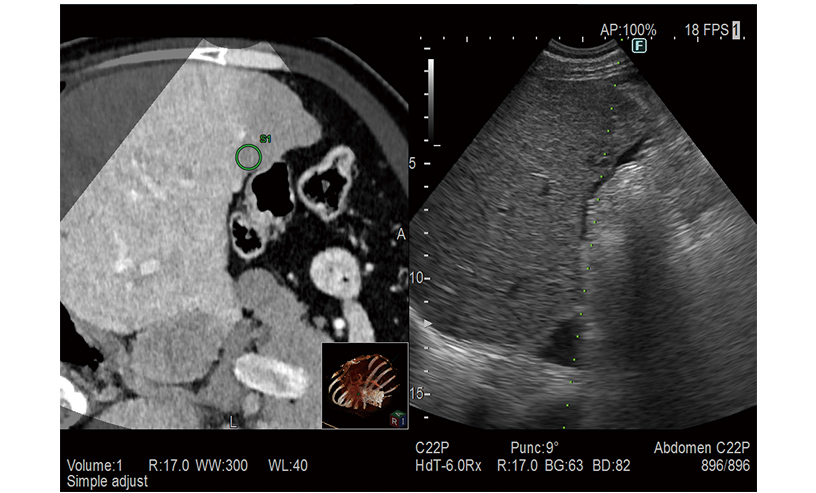
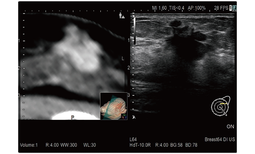
RVS is a function which fuses real-time ultrasound imaging with an MPR image created from the previously acquired CT, MRI or ultrasound volume data. RVS, a treatment supportting function by simultaneous display with other modalities, assists the positioning of treatment and the detection of small lesions that are difficult to find by ultrasound alone.
Provides simulation of single or multiple needle paths during navigation to a target with RVS. The positional relationship between the marked target and needle paths can be assessed in real time using the 3D body mark, reconstructed from the virtual CT volume data, with additional C-plane display orthogonal to the needle path.
A color map superimposed on the CT image simulates the distribution of the electric field (E-field) from the given location of multiple electrodes during RFA treatment. The simulation can be made with different positions of the multiple electrodes or additional ablation to determine the optimal arrangement. This flexibility in planning the needle path can bring improvement to the treatment technique.
*1 Option