


The content on this page is intended to healthcare professionals and equivalents.
Conventional CT examinations*1 required operators to manually adjust the height of the patient table and the starting position based on the target area after positioning the patient on the table.
As a result, there was variability in setup time and positioning depending on the operator's level.
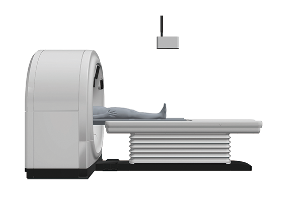
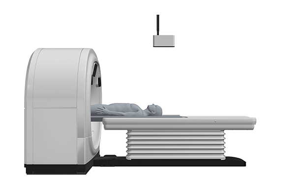
AutoPositioning*2,*3,*4 is a function utilizing deep learning that estimates the anatomical landmarks of the patient on the patient table in three dimensions from images captured by a ceiling-mounted 3D camera. The patient table automatically adjusts to the appropriate scanning height and position with a single button press. It supports a total of 14 different anatomical regions, including the head and chest.
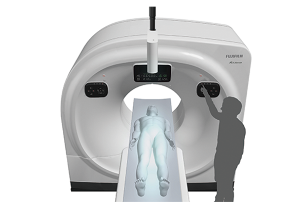
Supports 14 anatomical regions
The case of chest scanning
The case of chest and abdomen scanning
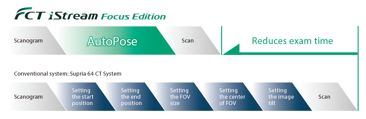
Conventional CT examinations*1 required operators to position the patient and take images called scannograms, on which operators would use a mouse to manually adjust the scanning range, field of view size, and tilt setting.
AutoPose, utilizes Deep Learning-based organ segmentation technology to automatically set the scanning range, field of view size, image tilt, and other parameters*5,*6. It supports a total of 14 anatomical regions, including the head and chest. Furthermore, when scanning the head, the iTilt function can be used to automatically create tilt images after scanning.
Supports 14 anatomical regions

RED:Scan range
Blue:Scan range + Margin
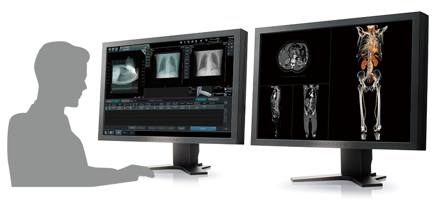
Fujifilm's image processing technology that developed over the years, now has been installed in the operator’s console, and the analysis function has been enhanced. Reconstructed images are simultaneously saved to the operator console and transferred to SYNAPSE 3D, which reduces the waiting time from scanning to starting analysis.
When used by one person
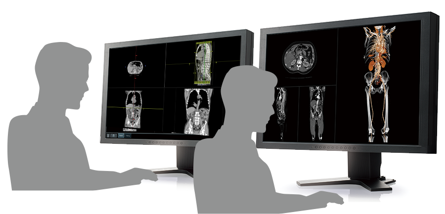
When used by two persons
The 3D image analysis system: SYNAPSE 3D*7, which extracts high-precision 3D images from CT and MRI images and performs analysis, has been further enhanced through Deep Learning by the medical IT technology brand: REiLI. By leveraging the vast amount of accumulated medical image data, the system now features enhanced automatic extraction functions for CT data. Leveraging image recognition technology, Fujifilm has achieved AI-era 3D analysis technology that supports team-based medical care.
Fujifilm's REiLI is leading the way to the forefront of team-based medical care.
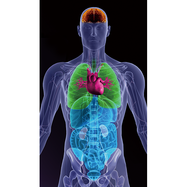
REiLI makes it happens to extract organs and simplify your work.
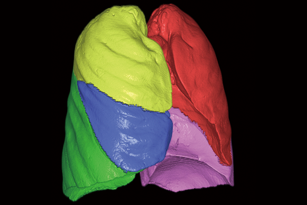
Lung lobe
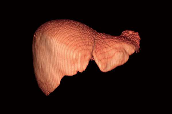
Liver
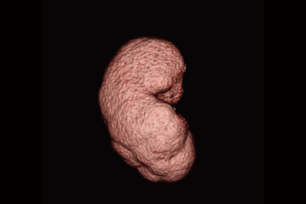
Kidney
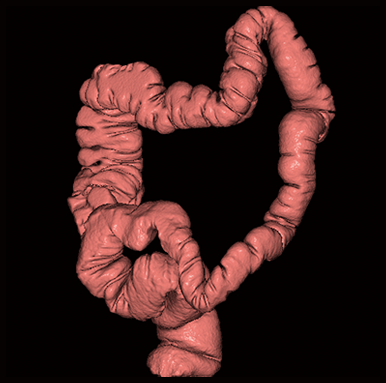
Colon
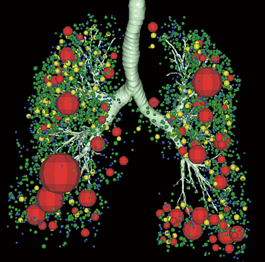
Lung Analysis
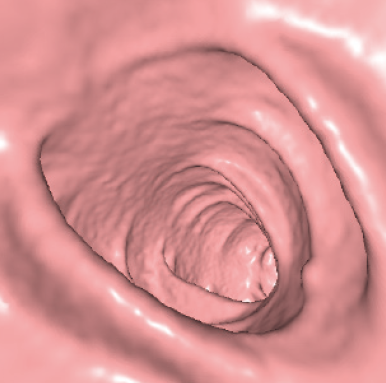
Virtual Endoscope
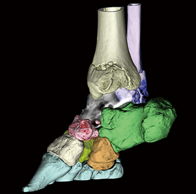
Orthopedics
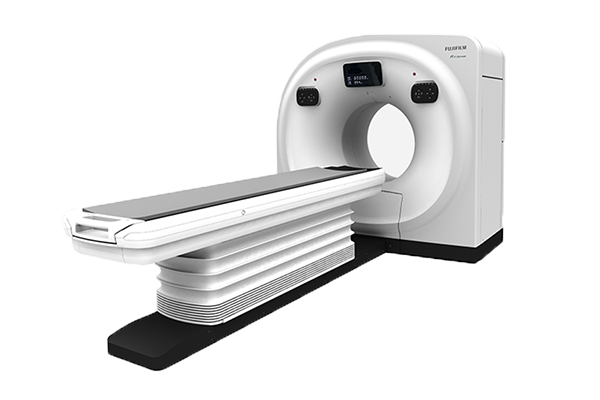
- *1 The case of Supria 64 CT System
- *2 Optional item
- *3 AutoPositioning was developed by utilizing Deep Learning, one of the AI technology. The performance and accuracy of the system do not automatically change after use.
- *4 Since AutoPositioning assists in moving the patient table for positioning, the operator needs to perform the final positioning and manually using a light localizer.
- *5 AutoPose was developed by utilizing Deep Learning, one of the AI technology. The performance and accuracy of the system do not automatically change after use.
- *6 The automatically set scan range must be checked, and the operator may adjust, if necessary.
- *7: SYNAPSE 3D is not included in the component of Whole body X ray CT System FCT iStream Focus Edition.