


The content on this page is intended to healthcare professionals and equivalents.
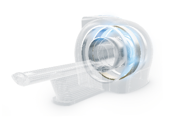
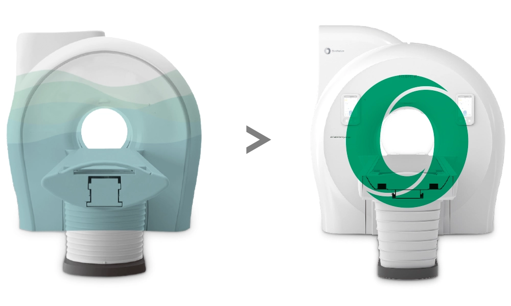
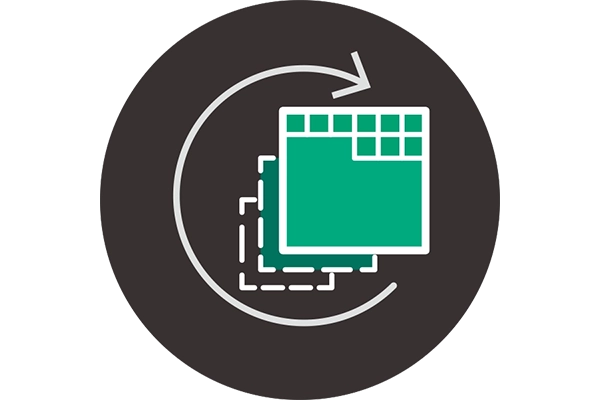
Introducing superconducting MRI to solve challenges in hospital management
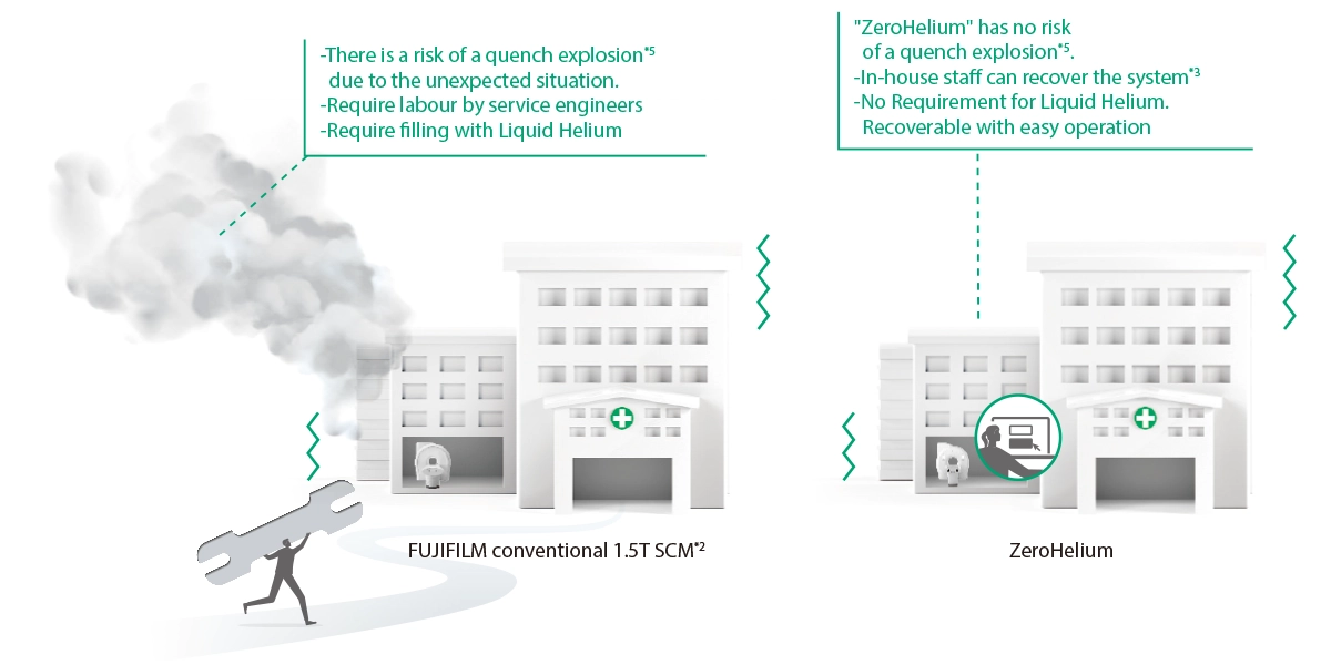
ZeroHelium technology cools the magnet without using liquid helium and significantly reduces recovery time and costs due to accidents and power failures from natural disasters.
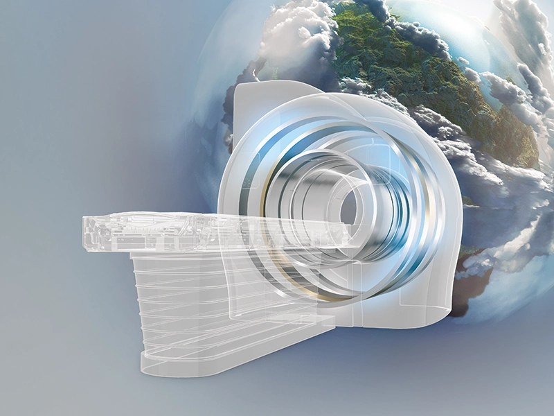
ECHELON Synergy ZeroHelium combines these advantages with high-quality imaging and workflows utilizing AI technology*1 to increase MR exam efficiency and patient satisfaction.
ECHELON Synergy ZeroHelium brings ease and efficiency to daily MRI examinations.
- *1 AI is utilized in the development process. The performance and accuracy of the device will not change automatically after installation.
Liquid Helium dependance to Zero
FUJIFILM conventional 1.5T SCM*2
Much of liquid helium is used.
ECHELON Synergy ZeroHelium
Zero liquid helium
- *2 ECHELON RX
“ZeroHelium” allows in-house staff *3 to ramp down and up the magnet in an emergency case*4.
That helps minimize the recovery time and cost called for recovery.
- *3 In some cases, service personnel may be required.
- *4 In cases where emergency ramp down is not required.
- *5 Represents the explosive release of helium when the superconducting state is lost.
Further Stable Operation with Three Values
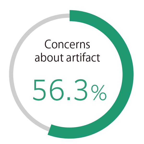
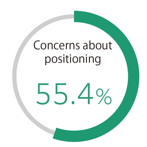
ECHELON Synergy ZeroHelium is equipped with technology designed to reduce concerns about prolonged MRI downtime caused by accidents or quenching, as well as minimize the risk of re-scanning due to artifacts or incorrect slice positioning. It aims to create an environment that reduces the physical and mental burdens on those involved in MRI examinations and more stable MRI operations.

- †4 "Questionnaire for technologists handling MRI," conducted in 2023 by mct, Inc.

REILI, FUJIFILM's medical Al technology brand, enables support for physicians in the diagnosis and streamlining of the workflow for diagnostic imaging by combining the image processing technology we have cultivated with the most advanced Al technology to realize improved medical care.

High quality and fast acquisition
Control image quality retrospectively
Save aborted scan
Monitoring cameras for patient safety and image quality
Automate complicated operations
Easy and flexible setting
- *6 Respondents who answered very concerned, concerned, or a little concerned
- *7 AI is used for development. The performance and accuracy of the equipment will not automatically change after installation

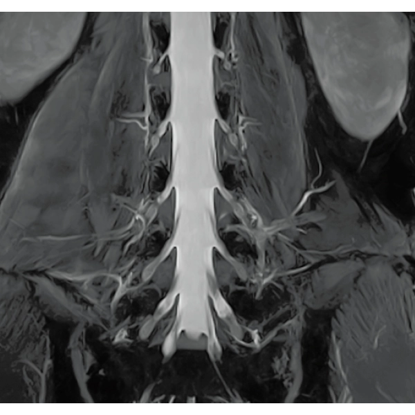
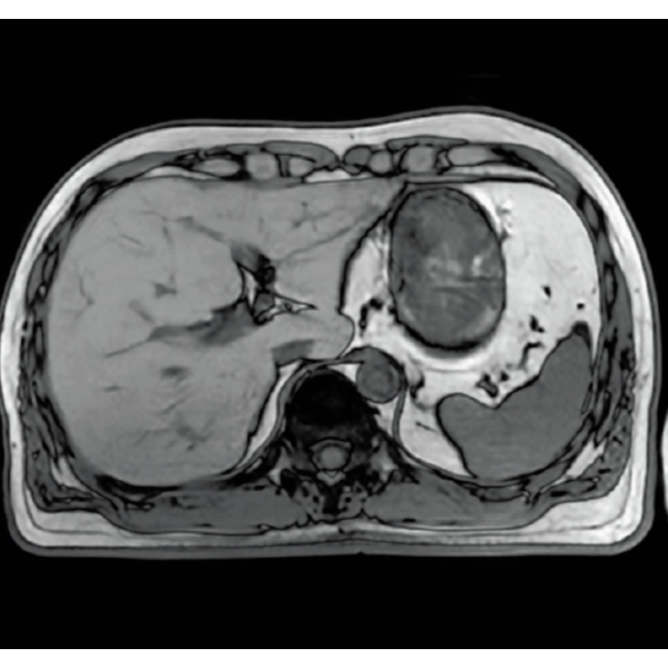
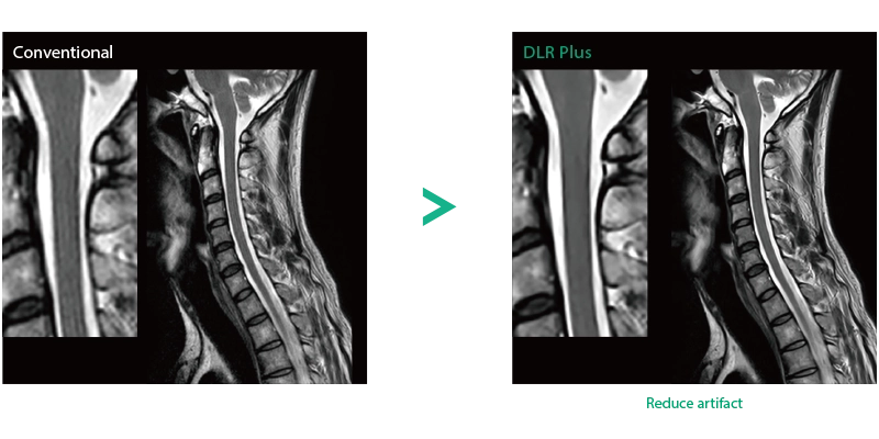
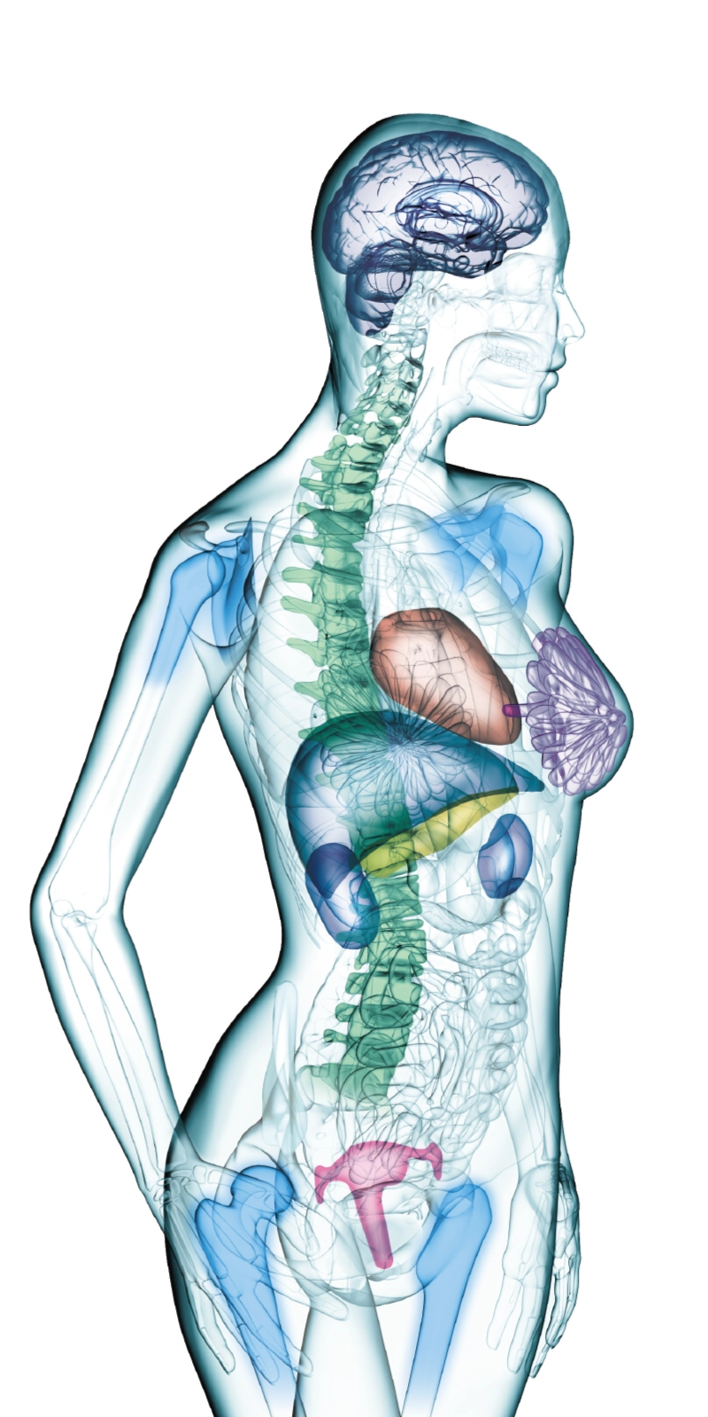
"DLR" is applicable to all body parts and promotes high speed imaging and diagnostic efficiency
IP-RAPID x DLR Plus can also shorten imaging time, allowing more images to be taken in the same examination time.
Additional imaging, such as different image types and cross sections, can be added to the conventional examination to increase the amount of information and make the diagnosis more reliable.
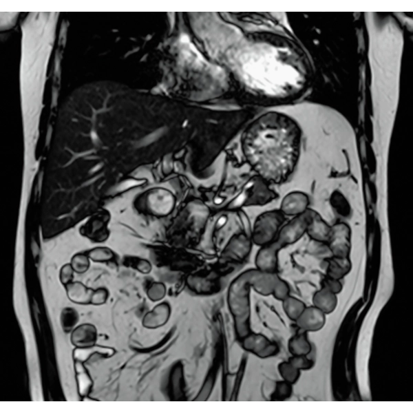
IP-RAPID x DLR Plus gives you the flexibility to shorten respiratory gated series or even replace them with breath-holds, depending on the patient’s situation. This gives you more options and a wider range of examinations to choose from.
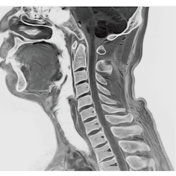
IP-RAPID x DLR Plus can be used for basic imaging such as VolumeScan, RadialScan, HalfScan, and many other imaging methods such as MultiContrastScan FatSep and DWI.
It can also be used with time-consuming scans such as Whole Body DWI and Whole Spine imaging, providing more detailed information in many areas than previously possible.
2DFSE
3DGrE Bone Imaging
3DPBSG
2DGrE
2DBASG

Imperfect imaging data to be better image quality through post-processing
By providing functions to remove and suppress artifacts in the imaging data and to generate reconstructed images from limited imaging data, it reduces the re-imaging rate and supports the realization of smooth MRI examinations.
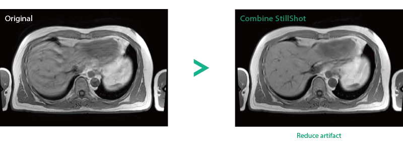
Reduce artifacts caused by patient movement, such as coughing or sneezing, by post reconstruction.
Improving SNR and reducing wrap-around artifact through post reconstruction.
Even if the examination is interrupted, the minimum necessary data can be reconstructed later.
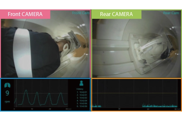
When motion artifacts occurs in the image due to coughing, sneezing, or involuntary movements, either or both the visual information acquired by monitoring cameras, Synergy Vision, and the intrabody information acquired by the navigator pulse can be used to provide an image with reduced artifacts. This reduces the re-imaging rate.
Body movements that affect image quality are detected from monitoring cameras based on thresholds derived for each body part.
Movements within the body are detected by navigator pulses. Body movements that significantly affect image quality are detected based on the error between pulses.
Deep Learning technology*8 enables image quality adjustment after imaging is complete. It optimize SNR and improve image sharpness by processing MRI signals in stages (k-space signal processing). This brings super-resolution and reduces truncation artifacts.
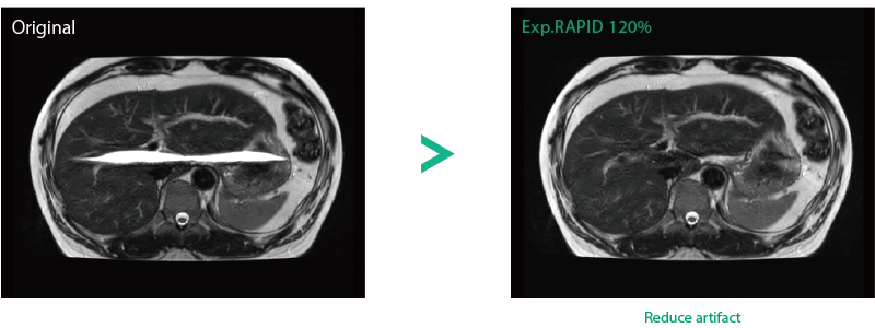
When the size in the phase encoding direction is incorrectly set , the signal outside the FOV will fold back and appear as artifacts in the image. Exp. RAPID reconstructs the image according to the specified FOV magnification ratio. Artifacts can be removed in post-processing.
- *8 Deep Learning is used for the development. The performance and accuracy of the device will not automatically change after installation
Series Save
When the scan has to be interrupted due to an emergency call by the patient, images can be reconstructed using the minimum amount of imaging data that has been obtained. This reduces the patient’s burden and operator's workload and contributes to the facilitation of operations.

Complex operations such as positioning are assisted
- Securing time to see the patient, enabling more attentive care.
- Supporting the task of viewing images, allowing time for checks and decision-making.
- Providing an environment to diagnose stress-free, minimizing variations in accuracy.
- Realizing operational efficiency and consistent quality from the perspective of looking at costs.
It provides a consistent and user-friendly operation environment, contributing to enhanced diagnostic quality and reduced workload.

AutoPose Cardiac GUI
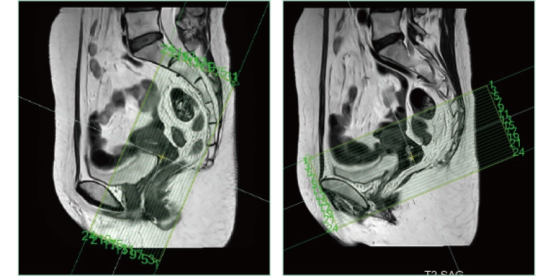
AutoPose FemalePelvis
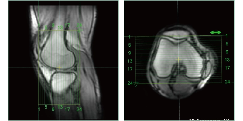
AutoPose Knee

Monitoring cameras are installed on both the front and rear sides of the bore. If the patient makes significant movements, it notifies the operator through screen alerts and sound. Additionally, the system can detect patient movements that may cause artifacts, such as coughing or sneezing.
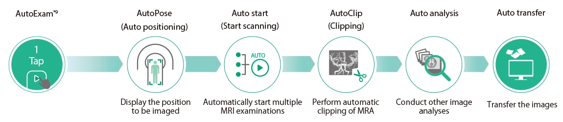
The slice line setting support function, "AutoPose," automatically sets the slice lines as soon as the scanogram is scanned.
After the completion of MRA imaging, it automatically performs clipping for cerebral MRA. The automatic clipping identifies the extraction range based on the characteristics of the head. Additional clipping can also be performed on the images after the automatic clipping process.
- *9 This system performs examinations and processing automatically but does not perform diagnoses automatically. Operator verification is required
The surface of parts likely to be in contact with the hands is designed to be easily cleanable by minimizing unevenness as much as possible. Antimicrobial films using FUJIFILM's antimi- crobial technology Hydro Ag+ are used for the gantry monitors, which are frequently touched. This ensures hygienic operation.

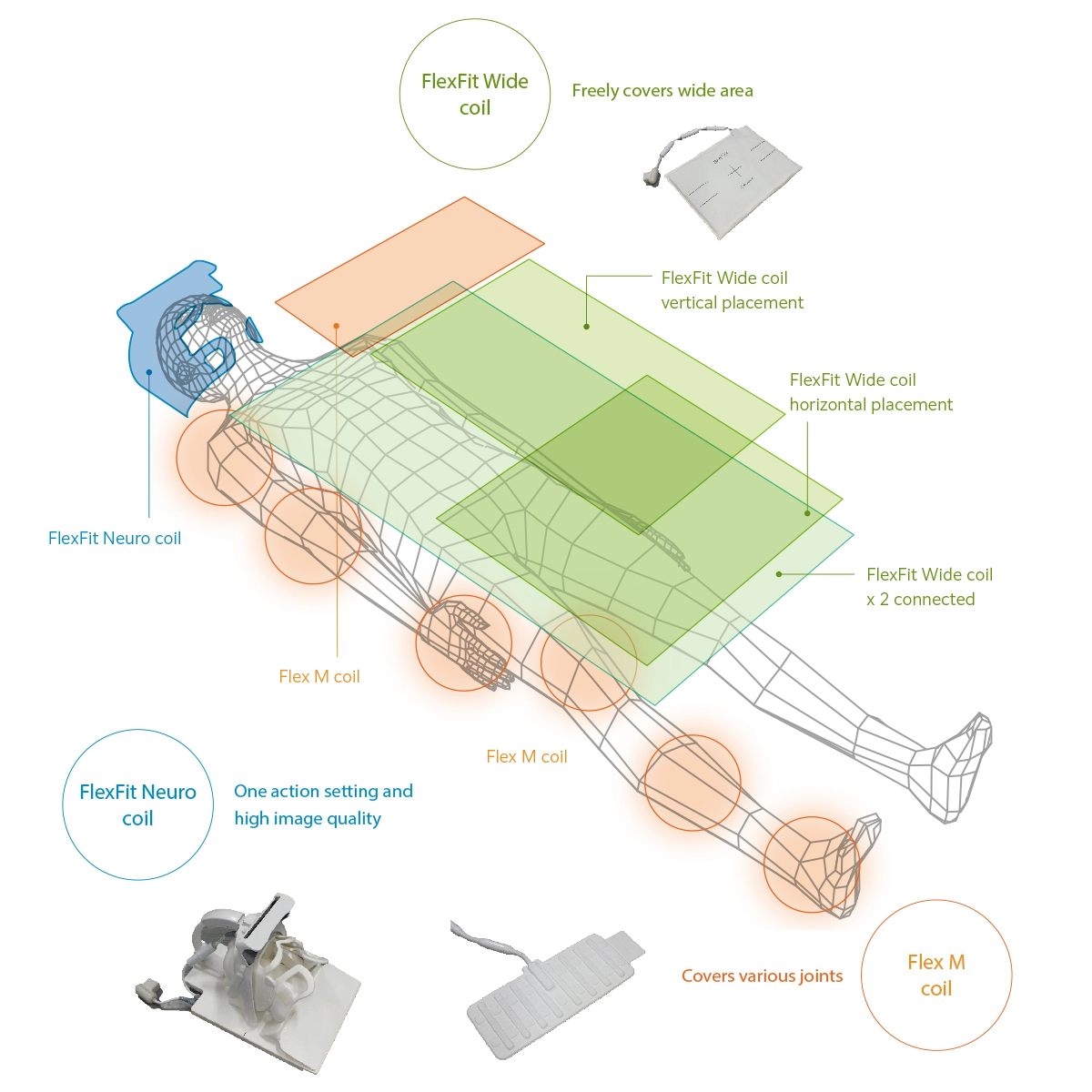
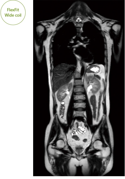
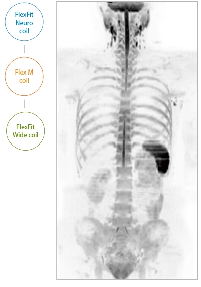
Wide bore and variety of coils to support comfortable examinations
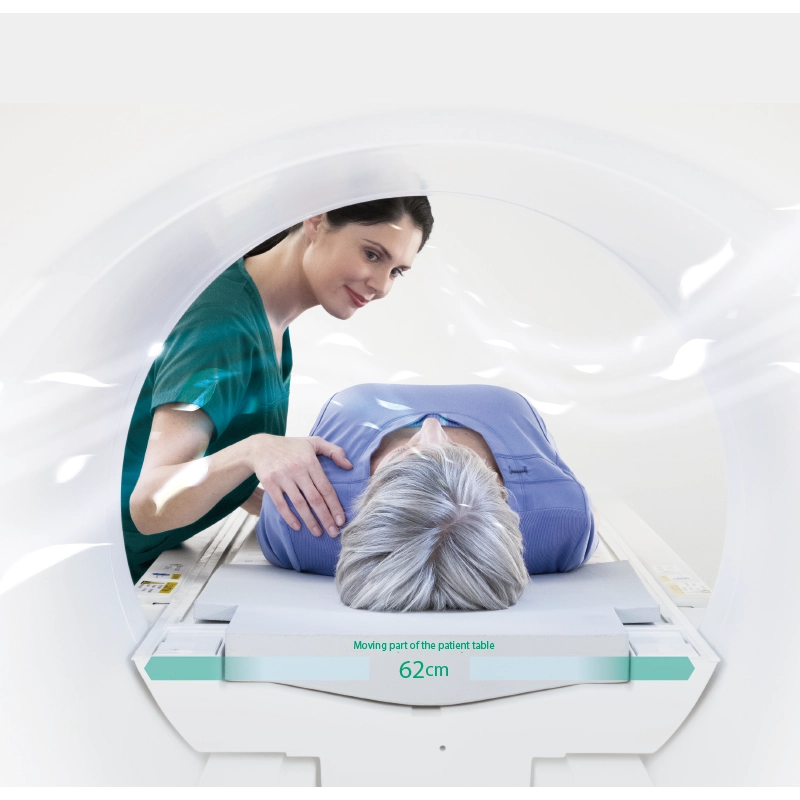
The 70cm diameter wide bore improves comfort for patients. To match the wide bore, we also paid close attention to the size of the patient table to ensure a space of 62 cm for the moving part of the patient table. This allows for quick adaption to imaging in a variety of patient positions.
- * Not applicable to all body positions and patients.
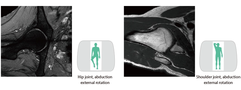
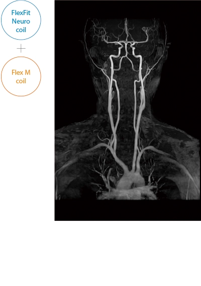
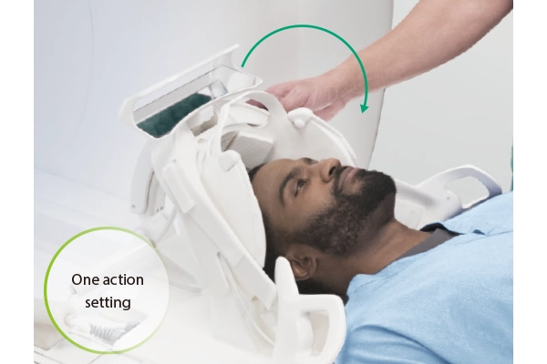
The ECHELON Synergy ZeroHelium is equipped with a flexible head and neck coil that allows one-action setup via sliding installation, as well as a Flex coil that provides wide and flexible coverage of the imaging area. This enables flexible adaptation to different part of body.
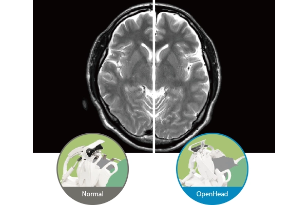
“OpenHead Mode“ which allows imaging without covering the anterior side of the coil over the patient’s face is equipped. By not obstructing the patient’s field of vision, this mode helps reduce the psychological stress associated with the imaging process.

“ZeroHelium” brings less concerns and burden both during and after installation
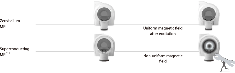
The ECHELON Synergy ZeroHelium provides advanced magnetic field control with its ZeroHelium magnet and ZeroHelium technology. Therefore, after demagnetization and excitation, MRI examination can be performed without confirmation by service personnel*10.
Before ramp down
After ramp up
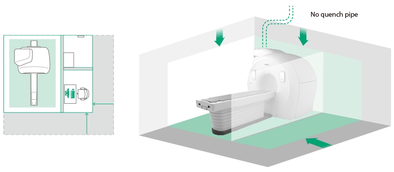
The ECHELON Synergy ZeroHelium does not require a quench pipe, which alleviates height installation constraints. In addition to the footprint of the equipment, the machine room area, including the ZeroHelium control unit, has been carefully designed to be equivalent to that of conventional wide-bore products.
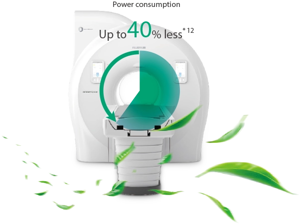
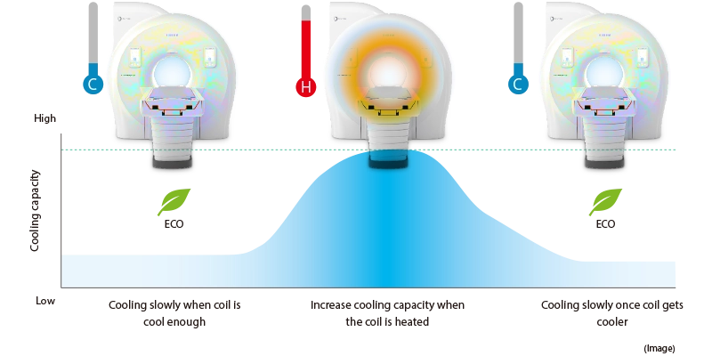
Monitors magnet temperature and varies cooling and power consumption based on need. Power consumption is reduced when cooling need is low and increases when temperature increases.
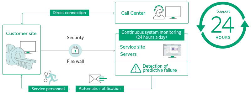
A predictive failure diagnosis service for superconducting MRI systems developed by utilizing the analysis of accumulated big data focusing on system operating status and FUJIFILM’s Predictive Failure Diagnosis Service. Utilizing 24-hour monitoring of the system status and predictive diagnosis by the service allowed us to optimize inspection and parts replacement cycles and improve system uptime.
- *10 Excluding quench and emergency demagnetization
- *11 ECHELON RX
- *12 According to our company's regulations. Varies depending on conditions of use