


The content on this page is intended to healthcare professionals and equivalents.
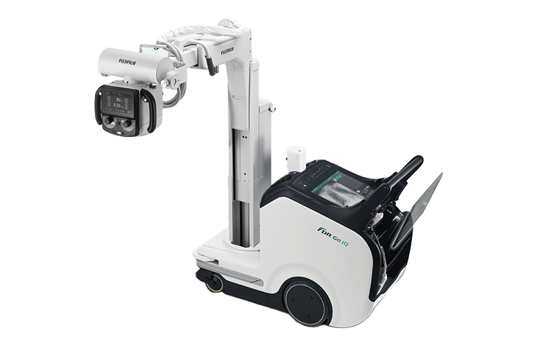
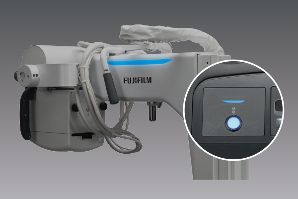
Equipped with a tube head monitor to display AI-estimated results*1*2
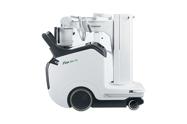
Collapsible column ensures forward visibility
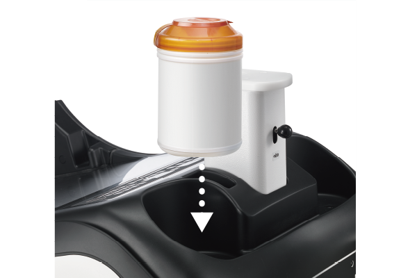
Built in smart charging for DR panel
Smooth maneuverability with quick-turning wheels
- *1 Based on Deep learning that is one of the AI technology
- *2 The system's performance and accuracy do not change automatically after installation
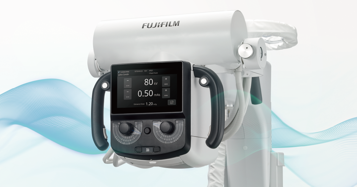
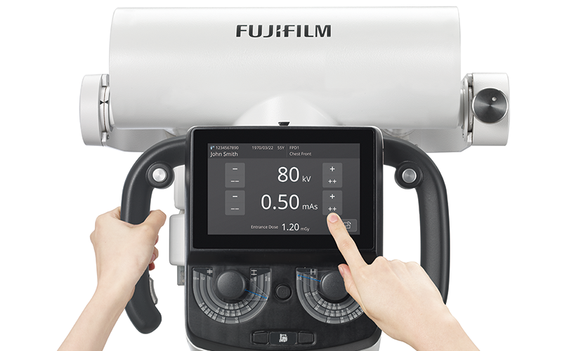
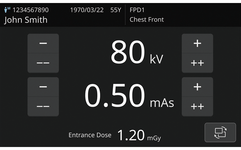
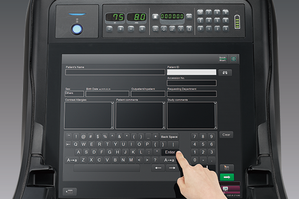
The touchscreen interface allows patient information and examination details to be displayed and exposure factors modified for an improved patient centric workflow.
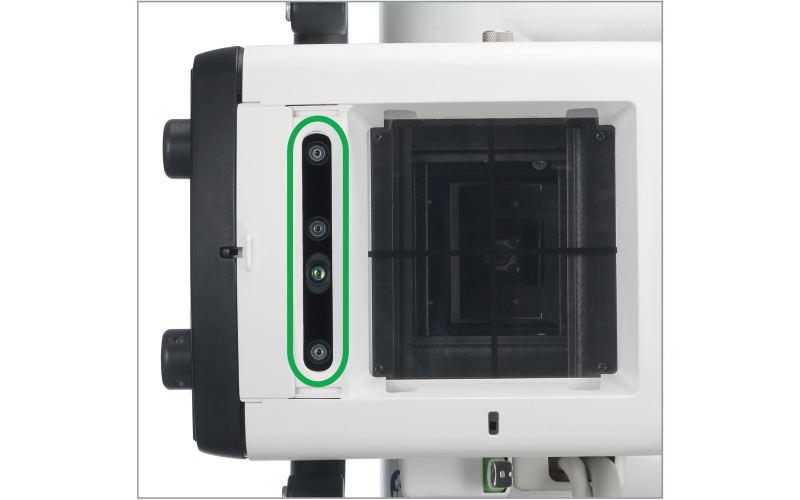
The camera built into the collimator provides live imaging display of the patient via the Tube head monitor for improved patient monitoring. The camera also allows detection of SID, angulation and alignment to enhance patient positioning.
- * Final visual confirmation is required to determine the irradiation field.
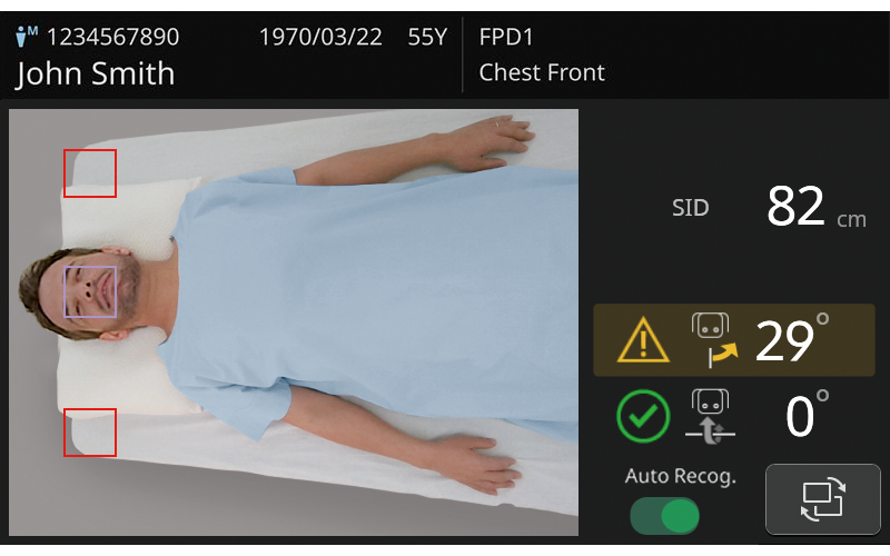
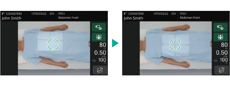
X-ray Centering Navi estimates the center position based on the camera image and highlights the center beam target for the body part selected.
- * Based on Deep learning that is one of the AI technology.
- * The system's performance and accuracy do not change automatically after installation.
- * Final visual confirmation is required to determine the irradiation field.
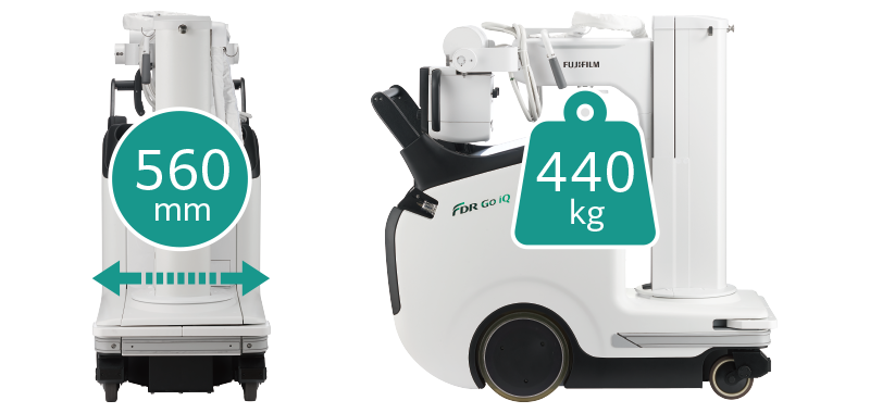
The slim and lightweight design provides excellent mobility even in tight spaces such as the bedside.
Built in smart charging for DR panel provides extended usability.
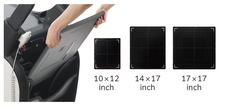
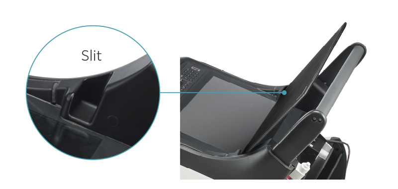
Cut-outs on the top of the main body are designed to hold the DR panel securely while adding sterile covers, cleaning or waiting to begin the examination.
A pocket is provided at the front left and right sides of the main unit, making it easy to carry wet tissues and accessories during imaging rounds.
The collapsible column provides extended visibility for greater safety when traveling
The large 19-inch touchscreen monitor and X-ray control panel, allows confident image verification and easy post processing.
LED lights on the side of the arm and next to the X-ray control panel, blink or change color to provide easy to identify system status notifications, such as Preparation for exposure and Charging mode or standby.
A high luminance green laser target, allows improved positioning in bright environments.
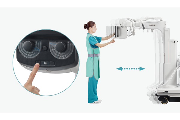
A release button has been added to the lower part of the handle. This helps operators with shorter stature position the tube without difficulty.
Controls on the collimator slowly move the system forward or backward, allowing precise bedside positioning without having to return to the drive handle.
The FDR Go iQ can be equipped with the external image processing kit, that provides the operating environment for AI-CAD. Analysis results can be shown on the console for improved POC clinical support.
AI-CAD processes images on the external image processing kit
The both Original and DICOM Secondary capture image generated by AI- CAD software are transferred to PACS
Read the Original image and check SC image generated by AI-CAD software on PACS
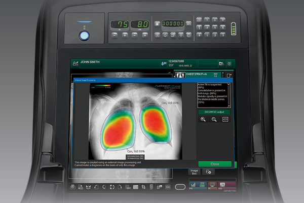
- * The console display is not for diagnosis purpose, it enables the user to check the result of software execution and send it to the PACS.
- * For the details on AI-CAD software, contact the software manufacturer.
- Model Name
FDR Go
- Manufacturer
SHIMADZU Corporation