The content on this page is intended to healthcare professionals and equivalents.
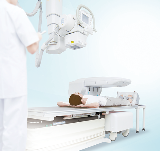
The ALPHYS LF, the flagship model of the ALPHYS bone densitometry brand, is the successor to the DCS-900 series, inheriting the same measurement accuracy and ease-of-use with a space-saving design that can be readily adapted.
Developed to be installed alongside existing X-ray systems and other imaging platforms.
Experience X-ray bone densitometry for the lumbar vertebra, femur and forearm with the “ALPHYS LF”, evolved from the DCS-900 series.


Conceived with ease-of-use in mind and with a compact size that allows installation alongside existing X-ray systems or other products already on the market.
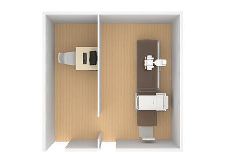
Can be combined with various types of X-ray imaging systems in a flexible arrangement to suit the room layout even in very limited spaces*1.
- *1 Please consult with our specialist staff for details.


Example of room layout with a ceiling mounted X-ray system


Example of room layout with wall mounted X-ray system


Example of room layout with a simple X-ray system
Can be installed with overhead or folding type imaging platforms*2.
- *2 Please consult with our specialist staff for details.


The large angle of tilt on the arm prevents interference with the X-ray tube.

Required footprint is approximately 0.68㎡, around 7% smaller than that of conventional system. Can be installed in small limited spaces.
The imaging platform at 55 cm high allows easy access for all patients.

- Approx. 55cm
The arm can be locked in the raised position.

- Release button
Button that releases in the locked position. - UNLOCK Switch
To release the arm when locked in the examination position.
Positioning guidelines with a scan area center line helps easy and accurate setting up to maintain patient’s comfort.
Reports can be prepared in various formats.
Uni-Report
The results of continuous measurements of multiple regions such as the lumbar and femur can be displayed on one report.
Parallel Report
Preparation of a basic report that enable the results to be clearly understood by all patients.
Features that enhance efficiency are provided at each step of the examination.
- POINT 1
The tilting arm and low height of the table result in easy patient access, rapid setting up and patient exit from the room. - POINT 2
The lumbar/femur holder and positioning guidelines result in quick and more accurate positioning. Use of the floating X-ray table supports smooth changes from a lumbar measurement to that of the femur. Additionally, the one-pass scan means a fast measurement time of about 40 seconds*3 for the lumbar and 20 seconds*3 for the femur, significantly reducing any patient discomfort. - POINT 3
Various report formats can be prepared.- *3 In standard mode

1. Corrects lordosis of the lumbar spine.

2. Corrects angle of anteversion.

* Forearm measurement kit is optional.
ALPHYS LF uses high quality image processing.

- High-resolution images achieved by using a multi-channel detector
Uses a 512-element detector to obtain high-precision imaging. - Easy Maintenance
Eliminates the routine phantom calibration required for maintenance. - BM Stabilizer
The BM stabilizer (bone mineral content stabilization mechanism) automatically adjusts the precision of each scan, controlling the accuracy. - High-output X-ray generator
Using the Tube voltage switching method to achieve high scanning speed. - Tube voltage switching method
Two different levels of X-ray energy can be obtained by switching the X-ray tube voltage. - One-pass Scanning
A wide-angle fan beam method suppresses beam distortion in order to provide bone density measurement values.
Operators can customize the image layout according to the exam type and their preferred format.

Customized layout

Standard layout
By referring to Context Help (the simple instruction manual), you can get guidance at any time.

Accurate analysis can be carried out with comparison to previously analyzed images using the same conditions.

When previous measurement data is available, the difference between the analyzed area to that of the previous exam is displayed on the screen.
- Pre-registration of patient information
- Metal Remove Function (artificial femoral head removal)
- CSV format compatible file output
- Analysis results display function


