


![[icon] Healthcare IT](https://asset.fujifilm.com/www/ph/files/2024-08/3514d54d9d3ec87c1d2d11b1b153b5ef/healthcare-it.svg)
![[icon] X-ray](https://asset.fujifilm.com/www/ph/files/2024-08/056e99b23ae4ef386de613c4ac064366/x-ray.svg)
![[icon] Ultrasound](https://asset.fujifilm.com/www/ph/files/2024-08/a1fb3473ad4be3b722188042b9ea0327/ultrasound.svg)
![[icon] Endoscopy](https://asset.fujifilm.com/www/ph/files/2024-08/5df55cd8106f48ed373ad5eb87f6fef4/endoscopy.svg)
![[icon] In Vitro Diagnostics](https://asset.fujifilm.com/www/ph/files/2024-08/cad75b9b4e5222c67aca1467675a9c22/ivd.svg)
![[icon] Veterinary Medicine](https://asset.fujifilm.com/www/ph/files/2024-08/6262ac987c5c5902046fe478868423e9/veterinary.svg)
![[icon] MRI and CT](https://asset.fujifilm.com/www/ph/files/2024-08/2956136b3bba4933876acdef77ab3c74/mri-and-ct.svg)
![[icon] Inkjet Solutions](https://asset.fujifilm.com/www/ph/files/2024-08/4d3a176eb96c6fcef8f0711241b5734d/inkjet-solutions.svg)
![[icon] Office Solutions](https://asset.fujifilm.com/www/ph/files/2024-08/0144d6162a335bcacc4b157636b9dea9/office.svg)
![[icon] Semiconductor Materials](https://asset.fujifilm.com/www/ph/files/2024-08/44ce61d4f214147c615c6fb4293b9056/semconductor-materials.svg)
![[icon] Graphic Arts & Printing](https://asset.fujifilm.com/www/ph/files/2024-08/96970efe3cca442b60b2e83620779aa9/graphic.svg)
![[icon] Photofinishing Products](https://asset.fujifilm.com/www/ph/files/2024-08/47fcfcf87a6be4d8010c1e1ed17cf986/photofinishing.svg)
![[icon] Optical Devices](https://asset.fujifilm.com/www/ph/files/2024-08/eb4f6d2e0535092537a53677f01074c7/optical-devices.svg)
![[icon] Cine & Broadcast Products](https://asset.fujifilm.com/www/ph/files/2024-08/caf6dd77bd95bc3084b20ec1408e0975/film-and-broadcast.svg)
![[icon] Security Protections](https://asset.fujifilm.com/www/ph/files/2024-08/7ebd1ccf4b89dbd1d69c50f9ab035cc5/security.svg)
![[icon] Manufacturing Process](https://asset.fujifilm.com/www/ph/files/2024-08/4a5936f0395fd84173c41992b9c96631/manufacturing-process.svg)
![[icon] Inspectional Products](https://asset.fujifilm.com/www/ph/files/2024-08/14eb3dc93110b75b0eda973e00156f79/inspection.svg)
![[icon] Data Management](https://asset.fujifilm.com/www/ph/files/2024-08/6ca23267b96be62c7fbdf7fc95aa9fa3/data-management.svg)
![[icon] Materials](https://asset.fujifilm.com/www/ph/files/2024-08/cea9ba81bb1d9c0dac3b392aaa1351eb/materials.svg)


The content on this page is intended to healthcare professionals and equivalents.
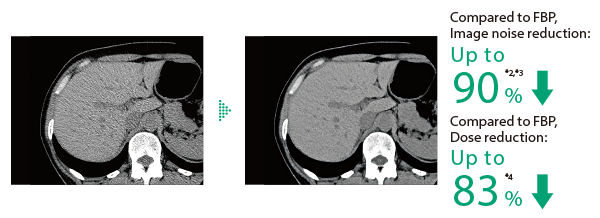
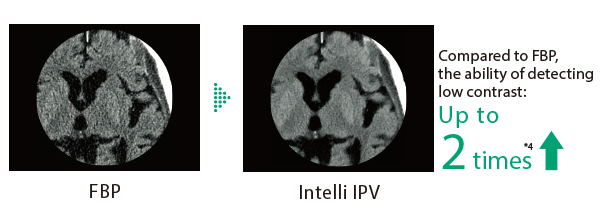
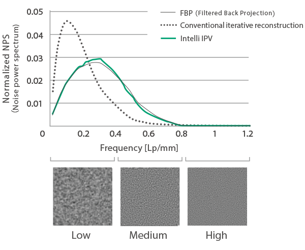
Intelli IPV is an image reconstruction developed using AI technology*1. By using images obtained through sufficient iterative processing as training data, the processing speed is accelerated. Based on the Visual Model developed by Fujifilm, image reconstruction processing starting from raw data reduces NPS: Noise Power Spectrum to FBP: Filtered Back Projection, maintaining image quality even at high noise reduction rates. It reduces image noise by up to 90%*2,*3 and radiation exposure by up to 83% *4. It also improves low contrast detection performance by up to 2times *4.
Visual Model controls image noise and image quality through repeated calculations based on statistical models, object models, and physical models.
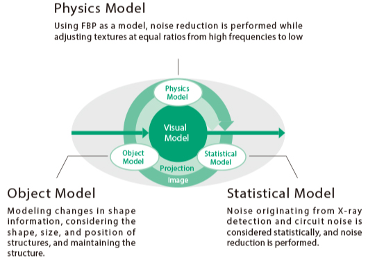
Intelli IPV has adjusted the texture across a ratio from high to low frequencies, bringing noise frequency characteristics that affect visibility as close as possible to FBP.
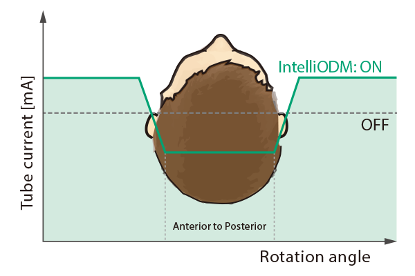
During head imaging, the X-ray irradiation from above the patient is reduced up to 30% by controlling the tube current. It reduces the exposure of radiation-sensitive areas such as the eye lens. This results in minimal changes of the image quality and allows the control range to be changed as needed.
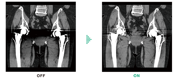
HiMAR Plus reduces metal artifacts using sequential approximation processing. It extracts metal parts,estimates artifacts, and then corrects them. The strength of the effect can be selected, allowing you to use it according to your purpose. HiMAR Plus can be used in combination with Intelli IPV.
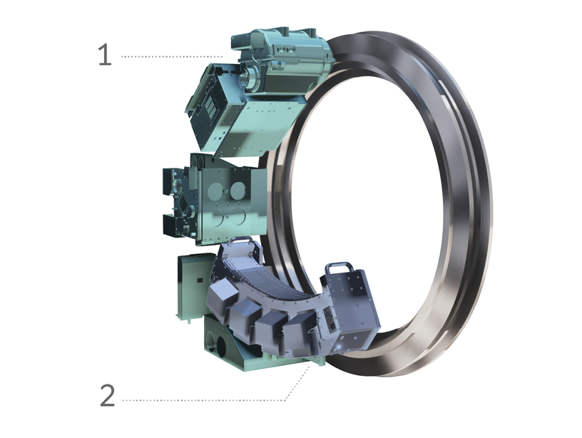
High-efficiency generator and 6MHU X-ray tube unit enable high output of 60 kW (Up to 670 mA). Also supports flexible scan conditions during low tube voltage scanning.
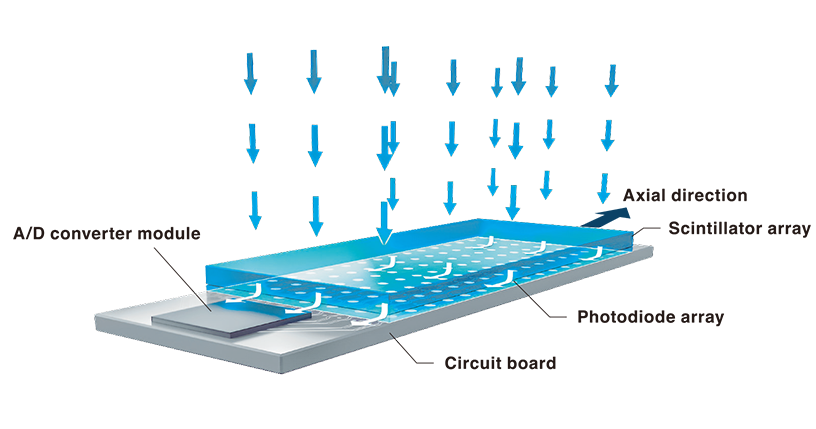
The optical system has been redesigned, and the detector has been upgraded. The detector employs MaxiLight technology.
By eliminating the need for analog wiring between bases, electrical noise is reduced, contributing to higher image quality.
- Sample clinical images from SCENARIA View CT System
- IPV stands for Iterative Progressive reconstruction with Visual modeling.
- *1 Intelli IPV was developed using Machine Learning, an AI technology. The performance and accuracy of the system do not automatically change after implementation.
- *2 It is obtained in the abdominal region.
- *3 Compared to FBP. It was measured using Intelli IPV intensity level Strong5 and tested to a water phantom. Depend on the clinical task, patient size, anatomic location, and clinical examination, the effect obtained may be smaller.
- *4 Compared to FBP. It was measured at 0.625 mm slice thickness using Intelli IPV intensity level Strong5 and tested to MITA CT IQ phantom: CCT189 which is made by Phantom Laboratory, using the model observer method results. Depending on the clinical task, patient size, anatomic location, and clinical examination, the effect obtained may be smaller.