


The content on this page is intended to healthcare professionals and equivalents.
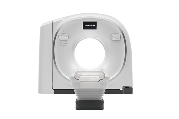
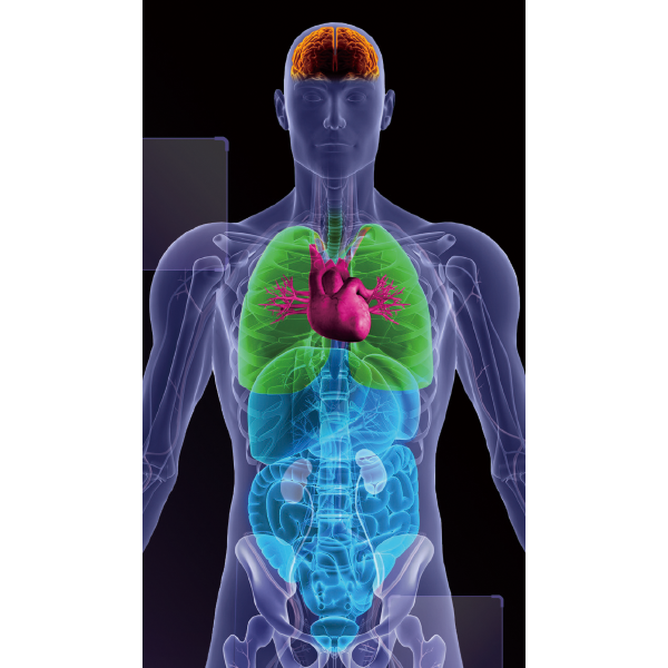
CT scans, which can examine the whole body in a short amount of time, are indispensable equipment in medical settings, and are used for diagnosis of various parts of the body, such as the head, heart, and abdomen.
We want to support people's healthy future with gentle examinations that are less of a burden on both the person being examined and the person doing the examination.
Fujifilm's experience and AI technology have led to further advances in CT.
REiLI, FUJIFILM’s medical AI technology brand, enables support for physicians in the diagnosis and streamlining of the workflow for diagnostic imaging by combining the image processing technology we have cultivated with the most advanced AI technology to realize improved medical care.
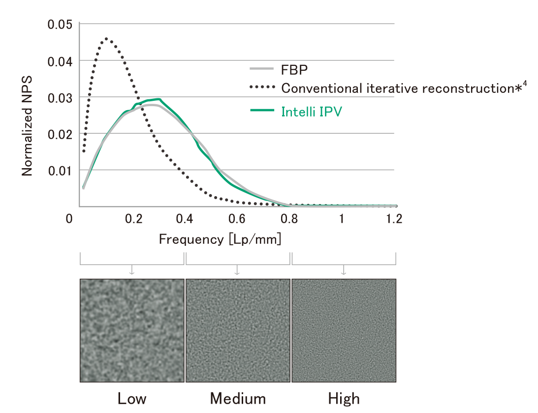
Intelli IPV is an image reconstruction technique developed with AI technology*1. Reconstruction processing has been speeded up by using images obtained through sufficient iterative processing as training data. Based on the Fujifilm's Visual Model, reconstruction processing using RawData brings the NPS (Noise Power Spectrum) closer to FBP (Filtered Back Projection) and keeps the image texture, even at a high noise reduction rate. It reduces image noise by up to 90%*2 and radiation exposure by up to 83%.*3 It also improves low contrast detectability by up to 2 times.*3
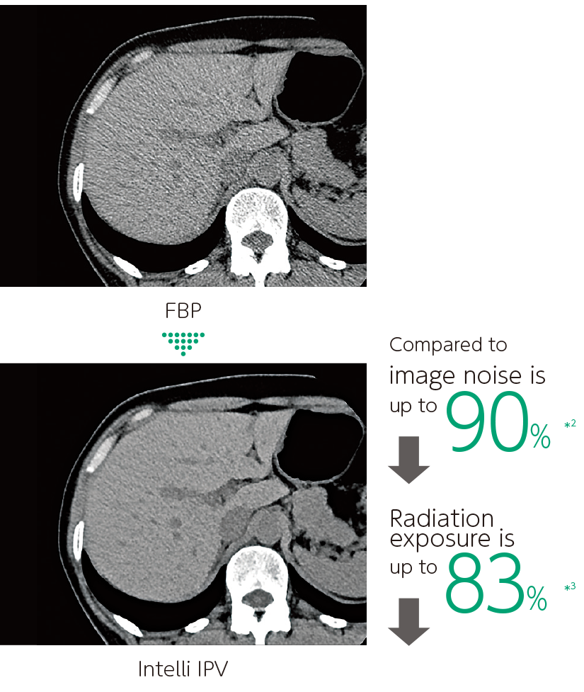
Achieved both radiation exposure reduction and visibility
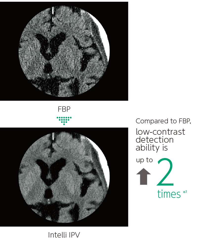
Improved low-contrast resolution
A technology to control image noise and image quality through iterative processing based on Statistical, Object, and Physical Models.
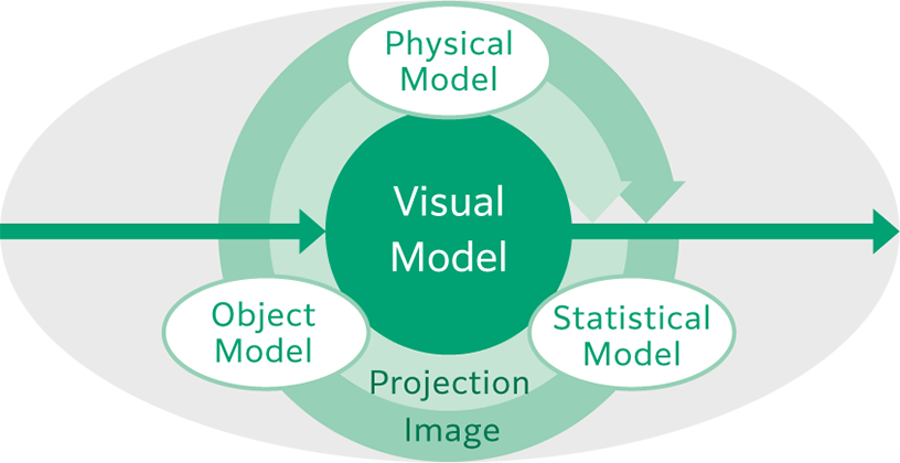
Reduces noise through statistical consideration of noise originating from X-ray detection and noise in circuit systems.
Models change in morphological information, and maintains structure considering shape, size, and position of the structure.
Modeled after FBP, adjusting texture in equal ratio from high to low frequencies while reducing noise to achieve a texture similar to that of FBP.
The noise frequency characteristics that affect visibility are now as close as possible to those of FBP while adjusting the texture in equal proportions from high to low frequencies.
- *1 Intelli IPV was developed using Machine Learning, an AI technology. The performance and accuracy of the system do not automatically change after use.
- *2 Compared to FBP. It was measured using Intelli IPV intensity level Strong5 and tested to a water phantom. Depending on the clinical task, patient size, anatomic location, and clinical examination, the effect obtained may be smaller.
- *3 Compared to FBP. It was measured at 0.625 mm slice thickness using Intelli IPV intensity level Strong5 and tested to MITA CT IQ phantom CCT189, Phantom Laboratory using the model observer method results. Depending on the clinical task, patient size, anatomic location, and clinical examination, the effect obtained may be smaller.
- *4 Our conventional iterative reconstruction is in our conventional CT system SCENARIA. IPV stands for Iterative Progressive reconstruction with Visual modeling.
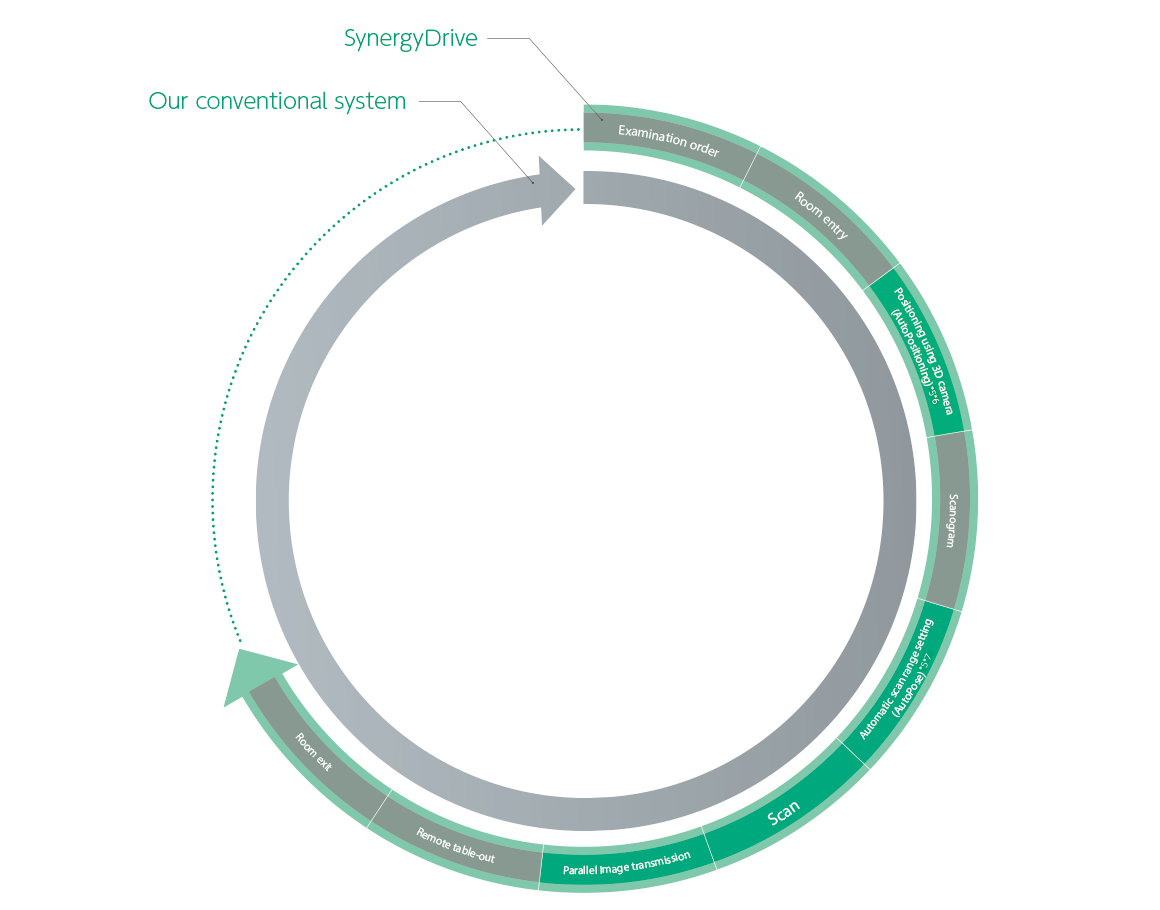
Speedy and Efficient Examinations for Everyone Involved in Clinical Practice
The workflow supporting functions, which was developed by utilizing AI technologies such as deep learning*5, help solve various issues in medical practice and contribute to increased efficiency and improved quality of medical care.
- *5 The performance and accuracy of the system do not automatically change after use.
- *6 AutoPositioning is an option. Since this function assists in moving the patient table for positioning, the operator needs to perform the final positioning and manually using a light localizer.
- *7 The scan range automatically calculated requires check and adjustment by the operator.
The patient table can move horizontally up to 200 mm, making it easier to position the scanned region in the center, even in cardiac or orthopedic areas, such as the shoulder. This is expected to improve examination efficiency.
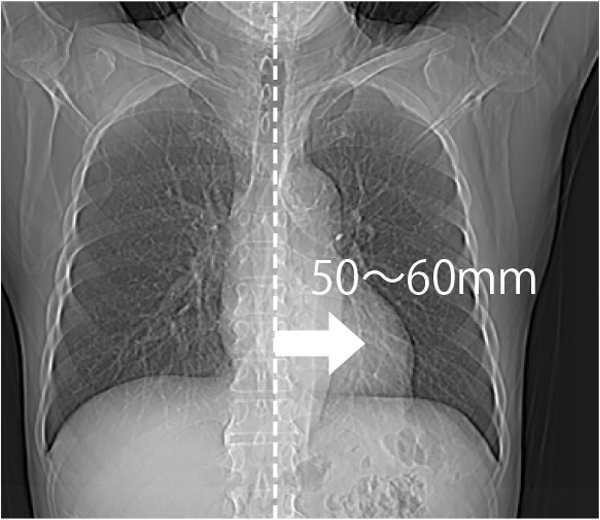
Positioning the heart near the center of the field of view
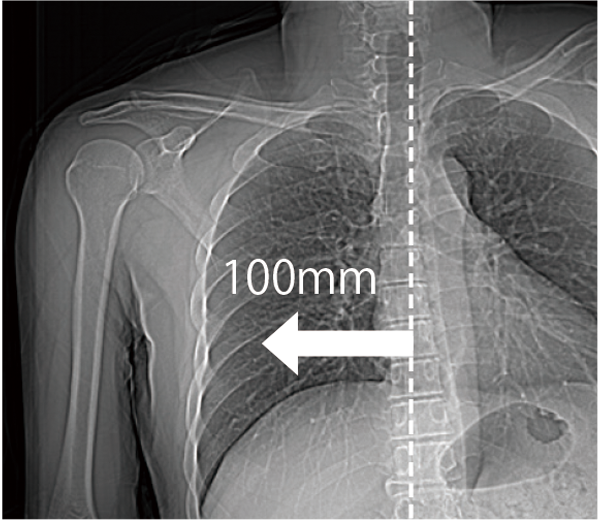
Positioning the extremities near the center of the field of view
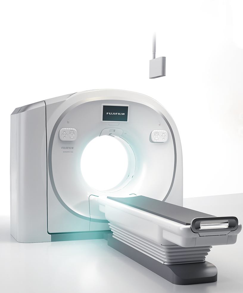
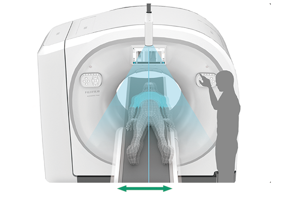
800mm wide gantry aperture with a smooth shape improve patient access.
Using technology developed utilizing deep learning*6, Using technology developed utilizing Deep Learning, the system recognizes the anatomical landmarks of the human body from 3D camera images, allowing you to set the bed in three dimensions (vertical, front-back, and left-right) with the touch of a one-button.It can be applied to 14 different types, with various patient orientations.In addition, the AutoPositioning video can be displayed on the Touch Vision on the front of the scanner or on the monitor of the Operating Console.

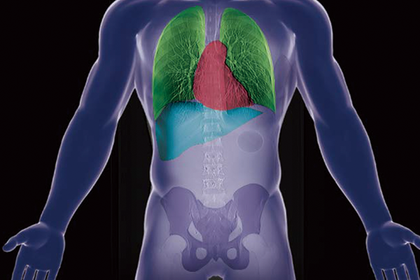
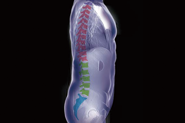
The captured scanogram image can be used to automatically set the scan range. This can be expected to be shorten the setting time. By using Fujifilm's automatic organ segmentation technology*7, which was developed utilizing deep learning, it contributes to the scan of a total of 14 types, including the head and chest. In addition, since the margin of the scan area can be set in advance, the scan area can be customized according to the operation of each facility. The operator can also check and adjust the automatically calculated scan area.
The image processing technology that Fujifilm has cultivated to date is also installed in the Operating Console of our CT system, enhancing the analysis functions. By simultaneously storing the reconstructed images in the Operating Console of our CT system and transferring them in parallel to SYNAPSE 3D, the waiting time from the end of the scan to the start of analysis can be shortened.
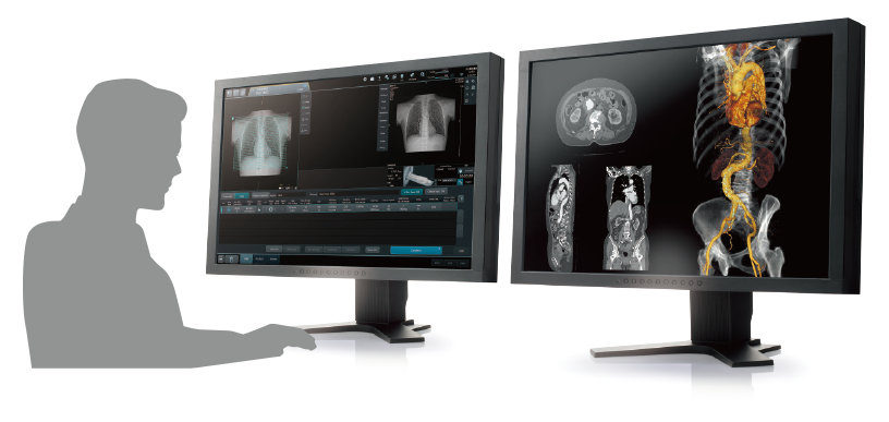
SYNAPSE 3D’s advanced image analysis technology aids clinical interpretation, reporting, and treatment planning. Especially,
automatic organ segmentation technology enables fast and effective workflow, powered by REiLI, FUJIFILM's AI brand. With
a series of high-tech applications developed in collaboration with clinical specialists, SYNAPSE 3D quickly and accurately
delivers imaging result that promotes effective care collaboration.
Based on the previously stored information, the areas recognized as blood vessels are extracted.
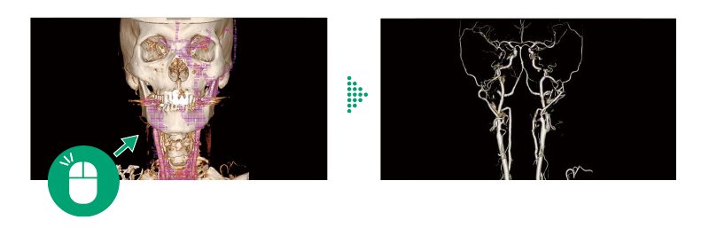
One-click operation to extract the areas that touch bones.
Vessels are extracted with one click by using image recognition technology.
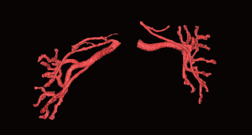
Renal Artery 3D
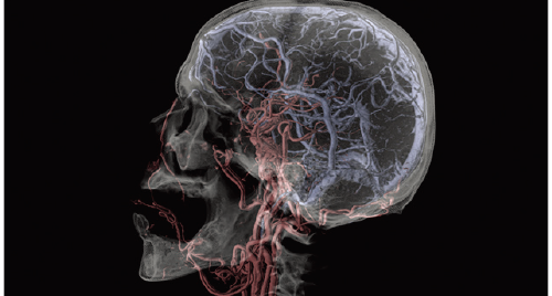
Cerebral Arteries and Vein separation
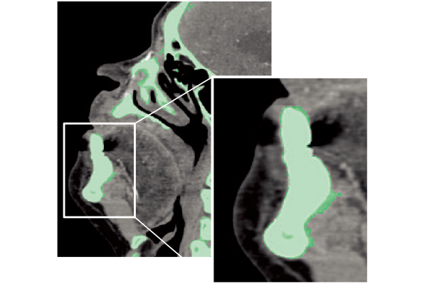
Bones are extracted or removed with one click based on the CT value and the shape of the region of interest recognized by REiLI technology.
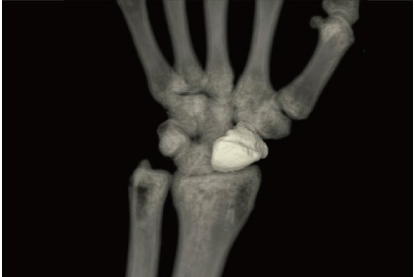
Carpal bones removal
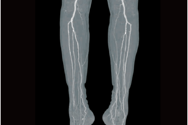
Lower extremity bones removal
Non-rigid registration enables SYNAPSE 3D to match an organ in images acquired at different phases and different time points.
Rigid Registration
Non-rigid Registration
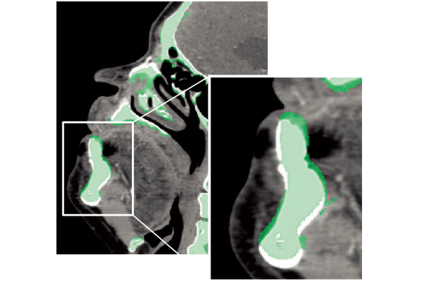
REiLI makes it happen to extract organs and simplify your work.
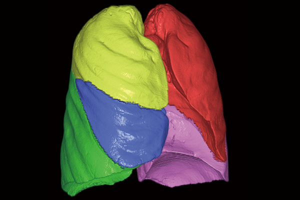
Lung lobe
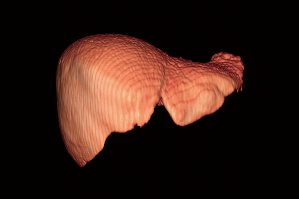
Liver
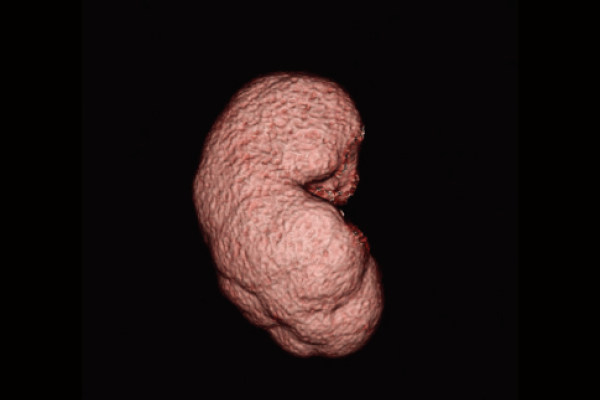
Kidney
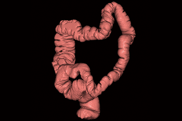
Colon
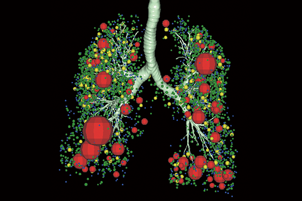
Lung Analysis
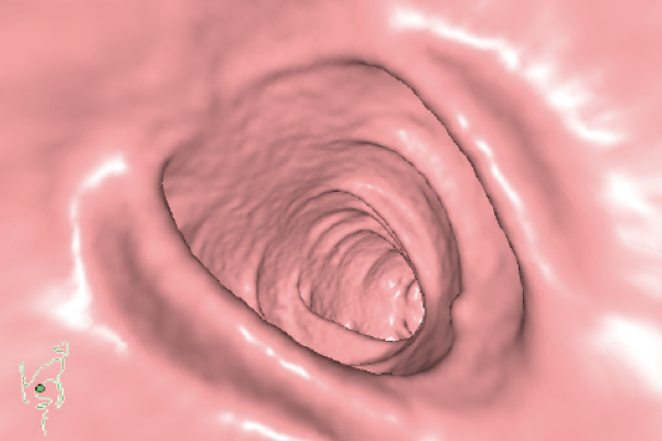
Virtual Endoscope
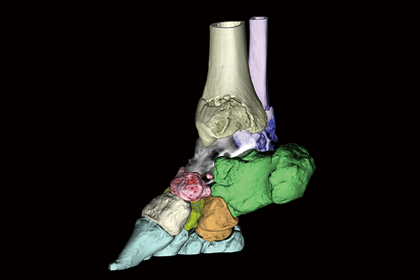
Orthopedics
