


The content on this page is intended to healthcare professionals and equivalents.
This is a function*1 that captures the heart rate range during breath-holding practice and automatically calculates the Scan parameters and image reconstruction phases based on ECG data. The operator can easily check whether the automatically calculated scan settings are appropriate or not with the displayed temporal resolution graph. The Parameter Setting Mode includes auto, semi-auto, and manual, which can be selected according to the purpose.
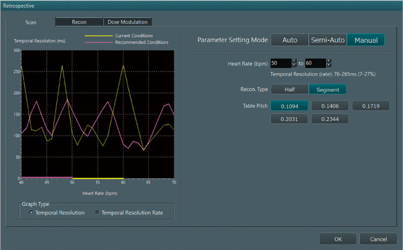
This function allows for an adjustment of the X-ray radiation dose based on ECG data. It reduces radiation in cardiac CT examinations by applying lower doses during non-target heart phases. The modulation of the tube current can be set up to two phases.

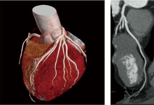
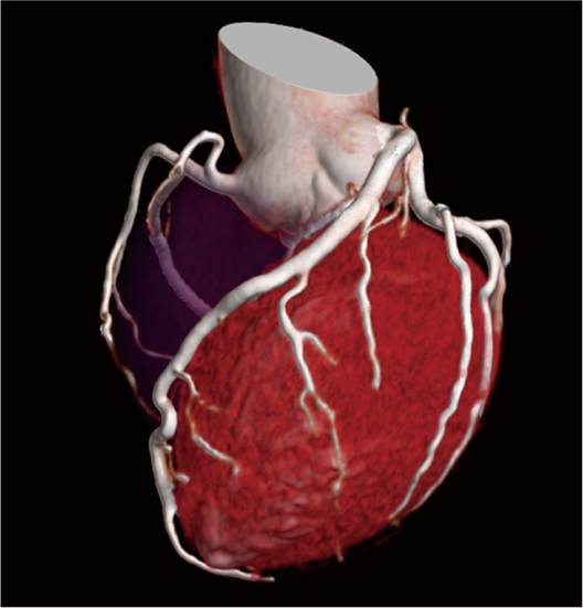
120kV, 0.35sec/rot, Beam Pitch 0.1719
Reconstructed cardiac phase 75%
This function automatically selects a cardiac phase with the least heart motion after cardiac CT scanning. It calculates the motion amount for each cardiac phase based on CT images and ECG data, and selects the appropriate phase with the least motion. CardioHarmony reduces the time required for finding the appropriate cardiac phase and image reconstruction necessary after cardiac CT imaging.

- *1 Checking setting, and adjustment by the operator required depending on the conditions used.
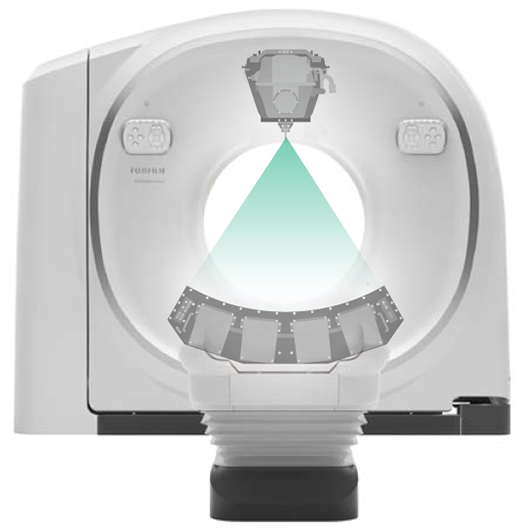
Breath-holding time during imaging and an increase in the amount of contrast agent can be a burden on the patient. SCENARIA View CT System is equipped with a high capacity 7.5 MHU X-ray tube assembly. It contributes to reducing the burden on patients by enabling examinations with the high output power required for high-speed imaging and low tube voltage imaging. Compared to conventional detectors*2, the reliability of the full-digital detector has been improved through the elimination of analog wiring between boards. It reduces electrical noise by up to 40% and power consumption by up to 45%.
HiMAR Plus(Right)
This technology reduces metal artifacts by applying iterative processing. Metallic parts are extracted to estimate the artifacts for correction. The effect level can be selected to meet your purpose. HiMAR Plus can be used in conjunction with Intelli IPV*3, an image reconstruction technology developed utilizing AI technology, to achieve noise reduction. It can also be applied not only during post reconstruction, but also multi reconstruction.
High-speed data sampling at 2,880 view/sec provides high resolution images with minimal artifacts at the edges. The rotation speed of 0.35 sec/rot used in cardiac CT can be applied to the whole body to shorten scanning time.
The On-time Standby function reduces power consumption by up to 47.0% compared to the unused state by controlling units built into the gantry. In addition, the Off-time mode function, which disconnects power while maintaining detector characteristics, reduces standby power consumption by up to 72.7% compared to the unused state.
Using the DICOM standard, it is possible to transfer dose information as a Structured Report(DICOM SR). Dose information can be transferred as a secondary capture from the CT console(Simple Dose Report).
- *2 Compared to the whole-body X-ray CT system SCENARIA.
- *3 IPV stands for Iterative Progressive reconstruction with Visual modeling. Intelli IPV was developed utilizing Machine Learning, an AI technology. The performance and accuracy of the system do not automatically change after use.
